
Human eye evolution may trace back to a one-eyed ancient ancestor
Lund University researchers say vertebrate eyes may have evolved from a median eye that also left a mark on the pineal gland.
By Priya Raghavan · Science Reporter
3 min read
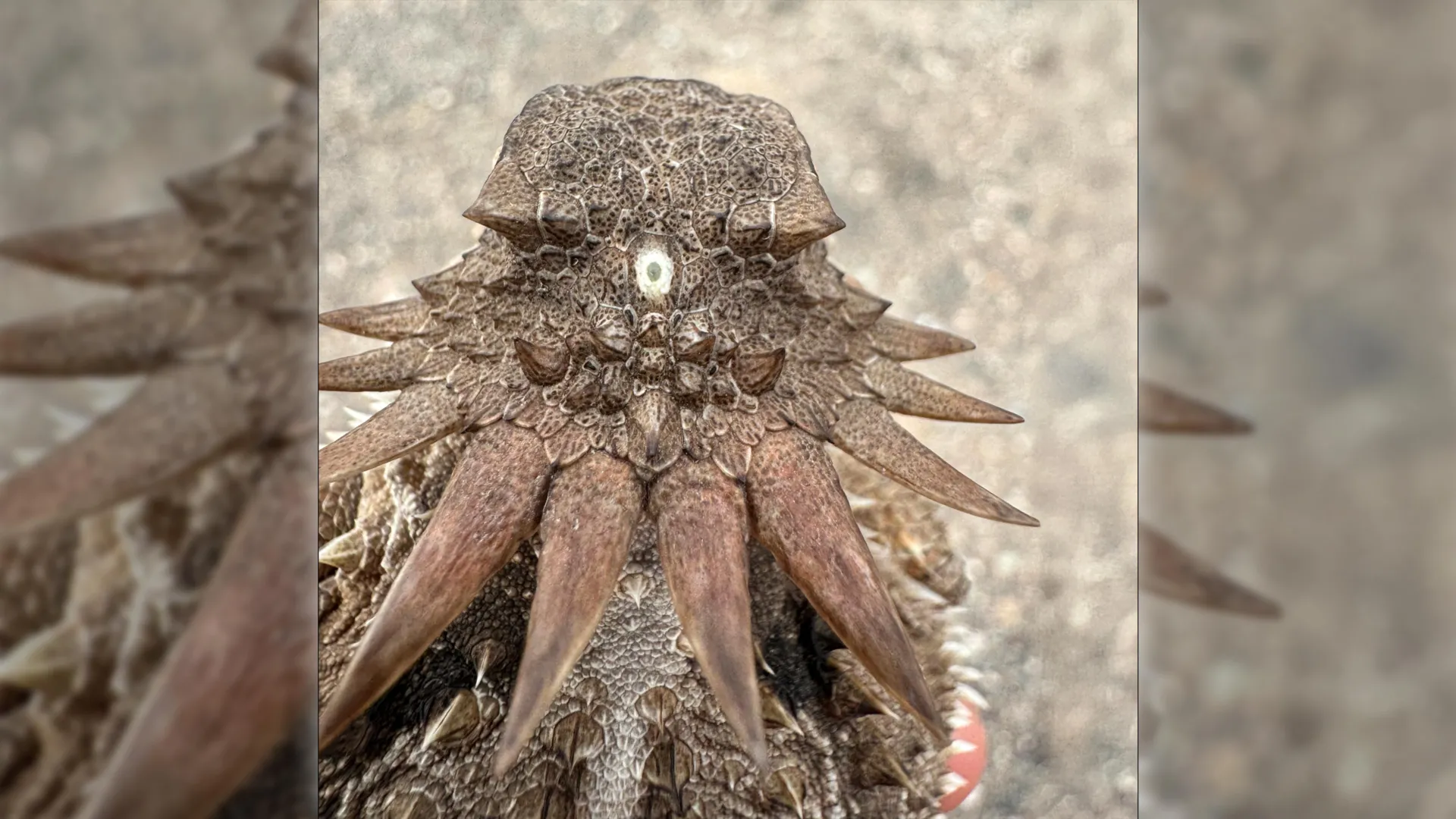
Human eye evolution may have taken an unusual route through a tiny, one-eyed ancestor that lived nearly 600 million years ago, according to researchers at Lund University and the University of Sussex. Their work argues that a central light-sensing organ in that animal helped set the stage for the paired eyes found in humans and other vertebrates.
The researchers also link that ancient median eye to the pineal gland, the small organ in the vertebrate brain involved in timing sleep with day and night. Lund University said the findings were published in Current Biology by George Kafetzis, Michael J. Bok, Tom Baden and Dan-Eric Nilsson.
Where did human eyes come from?
The study proposes that the earliest ancestor of vertebrates passed through a cyclops-like phase: a small ocean animal with a single eye-like structure on the top of its head. Lund University described the animal as worm-like and mostly stationary, feeding by filtering plankton from seawater.
According to Dan-Eric Nilsson, professor emeritus in sensory biology at Lund University, the organism appears to have had paired eyes, or at least paired groups of light-sensitive cells, earlier in its history. The researchers say those paired structures were later lost as the animal adopted a less active way of life and had less use for directional vision.
The animal retained a central patch of light-sensitive cells, however. That structure became a simple median eye, which likely helped it detect the difference between day and night and orient itself in the water, rather than form detailed images.
How the median eye became part of vertebrate vision
The researchers argue that descendants of this animal later became active swimmers again, creating renewed pressure for better vision. In their reconstruction, parts of the old median eye were repurposed over evolutionary time into a new pair of image-forming eyes.
That sequence would help explain a major difference between vertebrate eyes and the eyes of animals such as insects and squid, according to Nilsson. In vertebrates, the retina developed from brain tissue; in insects and squid, comparable eye structures arose from tissue on the sides of the head.
The retina is the light-sensitive layer at the back of the eye. It detects light and begins processing visual information before signals reach the brain, and the researchers say their work helps explain the origin of those retinal nerve circuits in vertebrates.
What does the pineal gland have to do with an ancient eye?
The pineal gland is a small organ inside the vertebrate brain that helps regulate daily biological rhythms. In humans, it produces melatonin, a hormone tied to sleep timing, with production rising and falling in relation to light and darkness.
Lund University said the researchers see the pineal gland as a surviving remnant of the ancestral median eye. Humans do not use it to see images, but the organ remains connected to the body’s response to daylight.
The conclusion comes from comparing light-detecting cells across animal groups, including where they occur in the body, how they work and how they connect with nerves and nearby tissues. The researchers say that comparison supports a model in which vertebrate eyes did not arise from the same structures that produced the eyes of insects, squid and other animals.
This story draws on original reporting from ScienceDaily.