
Study explains why H5N1 struck cow udders instead of lungs
University of Pittsburgh researchers say receptor patterns in mammary tissue explain the unusual course of H5N1 infections in dairy cattle.
By Priya Raghavan · Science Reporter
3 min read
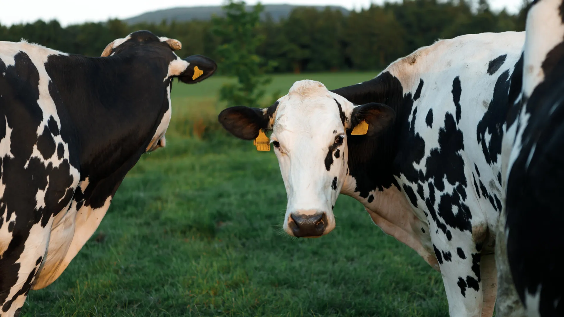
H5N1 bird flu caused severe udder infections in U.S. dairy cattle because the virus’s preferred cell receptors are concentrated in mammary tissue, according to University of Pittsburgh researchers. The finding may help scientists predict how the virus could behave if it reaches other animal species.
The work, published in Science Advances, examined why H5N1 looked different in cows than in other mammals after infections appeared in U.S. dairy herds in early 2024. According to Pitt Public Health, cattle developed serious mammary gland disease while their respiratory systems were largely spared.
The outbreak was first seen in dairy cattle in the Texas Panhandle, where animals suffered severe necrotizing mastitis, an inflammatory disease that damages mammary tissue, the university said. Veterinarians initially looked for common causes of mastitis in milk-producing animals, including bacterial pathogens.
Suresh Kuchipudi, chair of infectious diseases and microbiology at Pitt Public Health and senior author of the study, said the identification of bird flu as the cause surprised researchers and veterinarians. “We hadn’t even remotely considered that cattle could be a host for H5N1,” Kuchipudi said in the university’s announcement.
Before H5N1 was identified, the virus spread among herds and contaminated farm environments, according to the university. Kuchipudi said infected cows shed substantial amounts of virus into milk, raising concerns for farm workers and for animals exposed to raw milk, including domestic cats.
Pitt said Kuchipudi emphasized that pasteurization destroys the virus and that raw milk should be avoided. The university also cited previous work involving cases in which cats died after exposure.
The new study focused on influenza receptors, the cell-surface structures that allow flu viruses to attach and infect tissue. These receptors are glycans, sugar-based molecules that can vary by species and tissue type.
Earlier research from other groups had suggested that flu-related glycan receptors were present in cattle noses, tracheas and lungs, according to Pitt. That did not match the clinical pattern seen in infected cows, which were not showing the respiratory disease researchers expected.
To examine the mismatch, Kuchipudi worked with Lauren E. Pepi of Harvard Medical School, whose research includes glycomics, the study of glycan structures. The team used binding experiments, staining methods and high-resolution imaging to compare how H5N1 interacted with different cattle tissues.
The researchers found that H5N1 did not bind equally to all flu-related glycan receptors. According to the study, the virus attached to a specific subtype called N-linked sialic acid receptors.
Those receptors were widespread in udder tissue but nearly absent in airway tissue, Pitt said. The distribution helps explain why the virus caused severe mastitis in cattle rather than the lung-focused disease more often expected from influenza infections in mammals.
The researchers said the approach could be used to screen animals and tissues for possible susceptibility before unusual outbreaks become widespread. Kuchipudi said scientists could use receptor mapping to assess whether a species might show respiratory disease, mastitis or neurological disease, as his team has reported in cats.
Other authors on the study included researchers from Pitt, Pennsylvania State University, Harvard University and North Dakota State University. Pitt said the research was supported by Pitt Public Health and the U.S. Department of Agriculture’s National Institute of Food and Agriculture.
This story draws on original reporting from ScienceDaily.