
New MRI antenna sharpens images of the eye and deep brain
Researchers say a metamaterial-based MRI antenna could improve scans of hard-to-image anatomy without replacing existing scanners.
By Tom Brennan · Health & Medicine Correspondent
4 min read
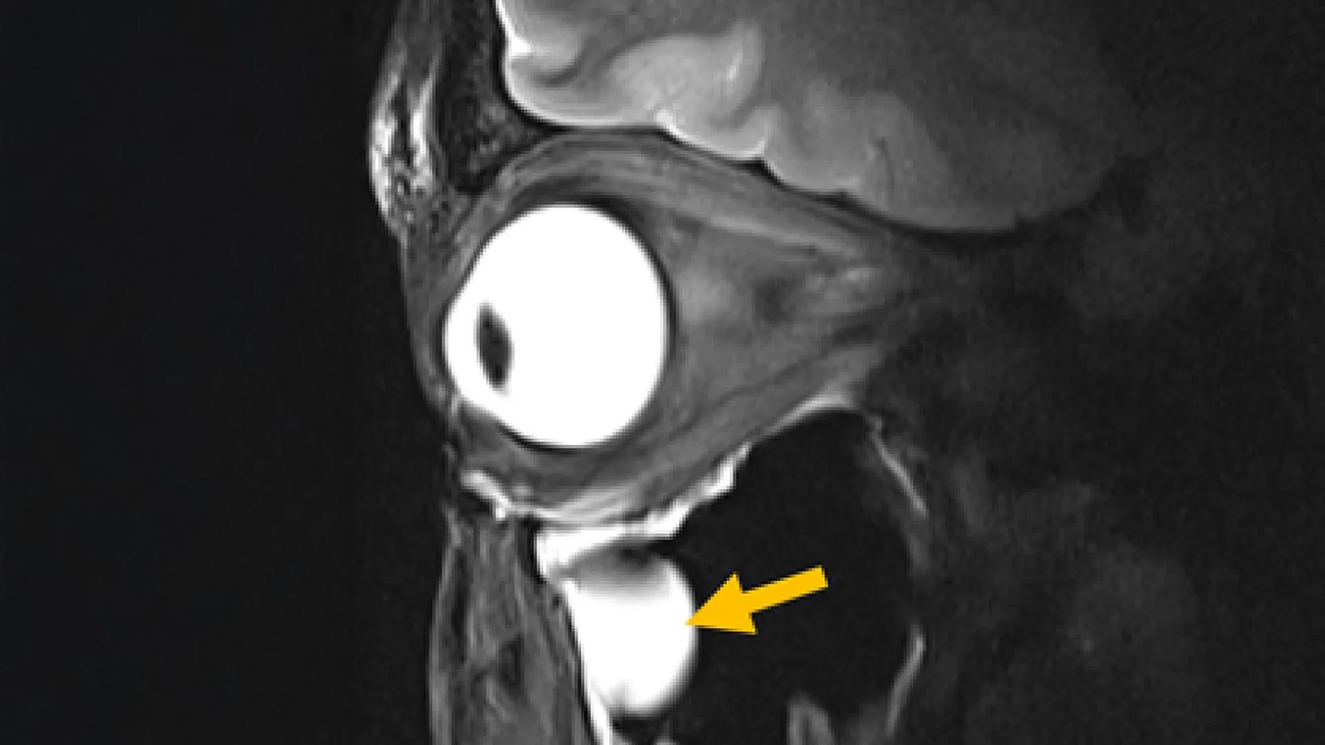
Researchers have developed an MRI antenna that they say can produce clearer images of the eye, orbit and parts of the brain while reducing scan time. The work could help doctors examine anatomy that conventional MRI hardware often struggles to capture in detail.
The device was designed by a team led by Nandita Saha, a doctoral researcher in Professor Thoralf Niendorf’s Experimental Ultrahigh Field Magnetic Resonance laboratory at the Max Delbrück Center for Molecular Medicine. The findings were published in Advanced Materials, according to the Max Delbrück Center.
MRI systems use radiofrequency signals and strong magnetic fields to generate images from the body’s tissues. The Max Delbrück Center said standard radiofrequency coils, the antennas that send and receive those signals, can have difficulty gathering strong signals from deep structures or complex regions such as the eye socket.
The research team addressed that limitation by building metamaterials into the antenna. Metamaterials are engineered structures designed to interact with electromagnetic waves in ways not usually found in natural materials, according to the Max Delbrück Center.
In tests, the antenna increased signals from selected tissues, improved image resolution and sharpness, and sped up data collection, the center said. The researchers tested the design on volunteers using a 7.0 Tesla MRI scanner to image the eye and orbit.
The image results included detailed views of anterior cranial structures, extraocular muscles and the optic nerve, according to the Max Delbrück Center. The center said the antenna can be added to existing MRI systems, which could avoid the need for replacement scanners.
Niendorf, senior author of the paper, said the work applied metamaterial concepts to better control radiofrequency fields. “This work shows a pathway toward faster, clearer MRI scans that could benefit patients in many clinical areas,” he said in a statement released by the center.
The project involved researchers in MRI physics, clinical ophthalmology and translational imaging from the Max Delbrück Center and Rostock University Medical Center. The Max Delbrück Center said the Rostock group is also helping evaluate the technology for possible clinical use.
Professor Oliver Stachs of University Medicine Rostock, a co-author, said the approach has clear relevance for ophthalmology because it may support high-resolution anatomical MRI of the eye. He said it could help researchers and clinicians study eye processes that have been difficult to access with previous imaging methods.
Saha said the team’s aim was to reconsider MRI hardware using modern antenna physics. According to the Max Delbrück Center, the same design principles might also be used to reduce unwanted heating near medical implants during MRI exams.
The center said the technology could also support MRI-guided cancer treatments by focusing radiofrequency energy more precisely, including in tumor hyperthermia or thermal tissue ablation. Those uses remain potential applications described by the researchers, not established clinical procedures for the new antenna.
Shorter scans could also make MRI exams more tolerable, the Max Delbrück Center said, especially when repeat imaging is needed because key details are hard to see. Because the antenna is compact and light, the researchers said it could be adapted for different body regions to improve comfort.
Niendorf said the design may be adjusted for MRI systems operating below or above 7.0 Tesla, according to the center. The team also sees possible uses in imaging organs beyond the eye, orbit and brain, as well as in monitoring metabolism or tracking how drugs move through the body.
The Max Delbrück Center said the approach may improve specialized MRI methods that image atoms other than hydrogen, including sodium and fluorine, by boosting signal strength and image quality. Dr. Ebba Beller of Rostock University Medical Center, a co-author, called the study a step toward next-generation MRI hardware.
The researchers are preparing larger clinical studies across multiple hospitals and are adapting the antenna for other organs, including the heart and kidneys, according to the Max Delbrück Center. The project was funded by the DFG as a joint collaboration between the Max Delbrück Center and the Medical University Rostock.
This story draws on original reporting from ScienceDaily.