
Vitamin A signal tied to formation of sharp central vision
Johns Hopkins researchers say lab-grown retinal tissue shows how fetal eyes form the cone pattern needed for sharp sight.
By Lucas Ferreira · Science & Environment Writer
3 min read
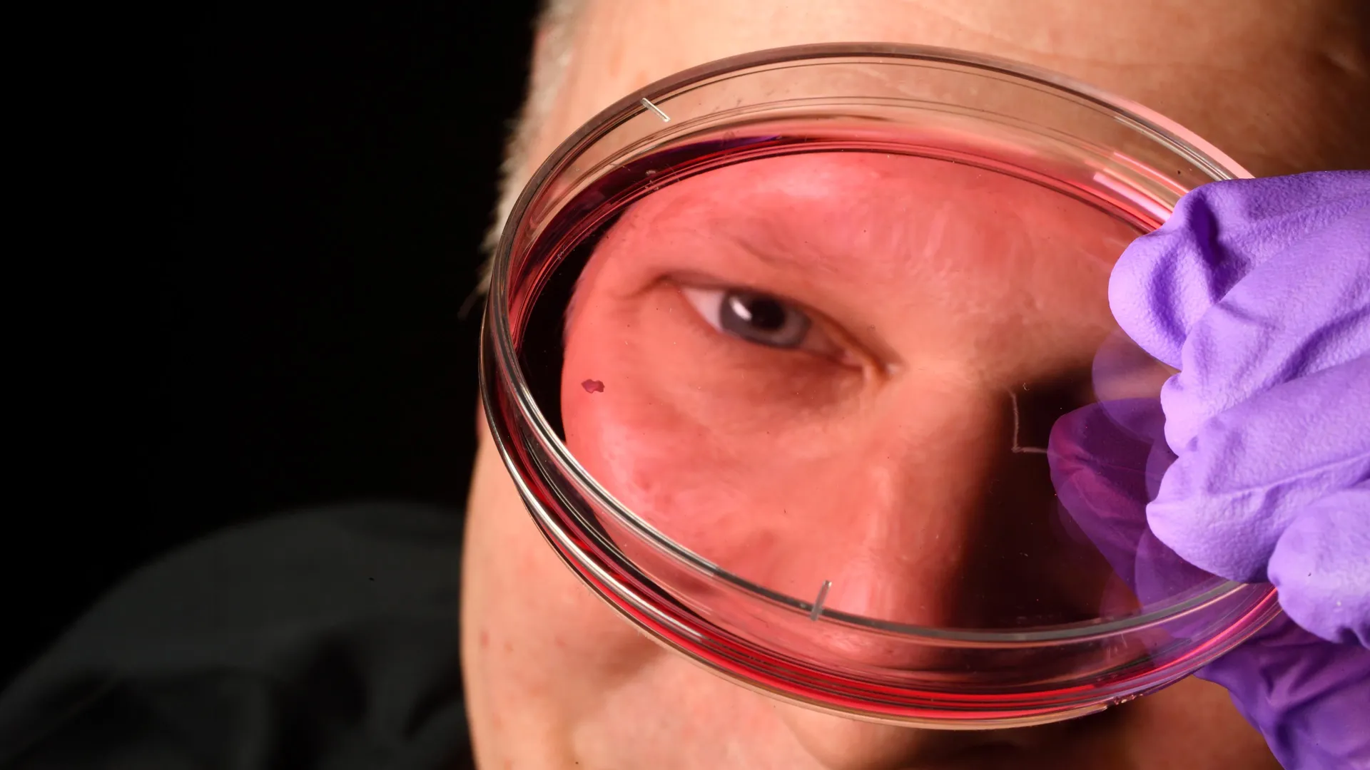
Johns Hopkins University researchers say they have identified a developmental process that helps build the part of the human eye responsible for the sharpest vision. The finding matters because it changes a long-running explanation of how key retinal cells form before birth and may help researchers improve future cell therapies for vision loss.
The study, published in the Proceedings of the National Academy of Sciences, examined lab-grown retinal organoids, according to Johns Hopkins. The team used the tissue models to study how the foveola, a tiny central region of the retina, develops its unusual pattern of light-sensing cells.
Robert J. Johnston Jr., an associate professor of biology at Johns Hopkins who led the work, said understanding the center of the retina is central to studying macular degeneration because that region is among the first affected by the disease. Johns Hopkins said the work may also inform research on glaucoma and other conditions that damage sight.
How the central retina takes shape
The research focused on cone photoreceptors, the cells that support daytime and color vision. According to Johns Hopkins, these cells develop into blue, green or red cones, each responding to different wavelengths of light.
The foveola is small, but Johns Hopkins said it accounts for about half of human visual perception. The university said the region differs from the rest of the retina because it contains red and green cones, while blue cones are absent there in the mature pattern.
Johnston’s team tracked the organoids over several months to follow how that pattern forms. According to the study summary from Johns Hopkins, a small number of blue cones appear in the developing foveola during weeks 10 to 12 of fetal development, but by week 14 those cells have become red and green cones.
The researchers linked that switch to two signals, Johns Hopkins said. Retinoic acid, a molecule derived from vitamin A, is broken down in a way that reduces the production of new blue cones, and thyroid hormones then push remaining blue cones to take on red and green identities.
A revised model for cone cells
The finding challenges a roughly 30-year-old model in vision research, according to Johnston. That earlier explanation held that blue cones formed in the central retina and later moved away from the area.
Johnston said the new data support a different mechanism in which cells stay in place and change type over time. He also said the team cannot fully rule out the older model, according to Johns Hopkins.
Johns Hopkins said the question has been difficult to study because common laboratory animals, including mice and fish, do not develop the same human cone arrangement. The organoids gave the team a way to observe human-like retinal development in the lab.
Possible use in future therapies
The researchers said the work could help improve retinal organoids so they more closely match human retinal function. Johns Hopkins said better models may aid efforts to produce healthy photoreceptors for possible cell replacement therapies.
Katarzyna A. Hussey, a study author who is now a molecular and cell biologist at CiRC Biosciences in Chicago, said the long-term goal is to make tailored populations of photoreceptors that could be introduced into the eye. She said such work would require further safety and efficacy studies before any clinical use, according to Johns Hopkins.
The journal reference lists Hussey, Kiara C. Eldred, Brian Guy, Clayton P. Santiago, Jingliang Simon Zhang, Ian Glass, Thomas A. Reh, Seth Blackshaw, Loyal A. Goff and Johnston as authors of the PNAS paper.
This story draws on original reporting from ScienceDaily.