
Scientists map eye molecules that help humans see color
Researchers report the atomic structures of human cone opsins, clarifying how eye cells convert red, green and blue light into signals.
By Priya Raghavan · Science Reporter
3 min read
A research team in China, Germany and Australia has resolved the atomic structures of human cone opsins, the light-sensitive molecules central to color vision. The work, reported by the Australian National University and published in Science, gives researchers a molecular-level view of how the eye turns different colors of light into signals for the brain.
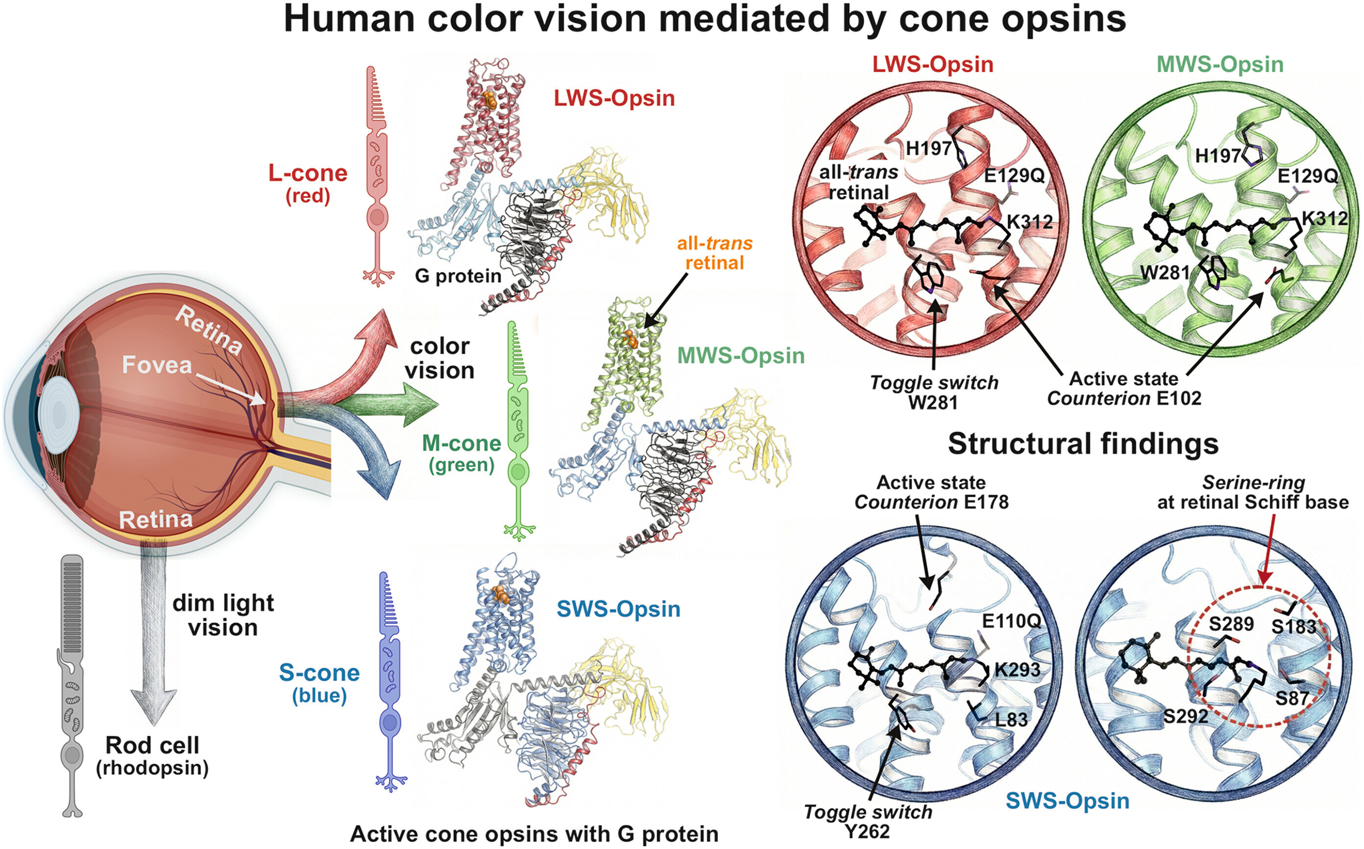
According to ANU, the study examined the three cone opsins found in retinal cone photoreceptor cells. Those molecules are associated with red-, green- and blue-sensitive vision, and their relative activation is a main factor in how people perceive color.
ANU researcher Emeritus Professor Trevor Lamb, who worked on interpreting the molecules’ role in the eye, said understanding color detection requires knowing the exact structure of the eye’s light-sensitive molecules. The university said Lamb is based at the John Curtin School of Medical Research.
The paper, by Qi Peng and colleagues, is titled “Cryo–electron microscopy structures of human cone visual pigments” and appears in Science. ANU said the structures show the cone opsins in their light-activated state, when they begin the process that leads to visual signals being sent onward to the brain.
How cone opsins tune light
ANU said each of the three cone opsins uses the same vitamin A-derived light-sensitive molecule, retinaldehyde. The difference is in how each opsin binds to retinaldehyde, allowing the molecules to respond to different wavelengths of light.
According to Lamb, the study explains how each cone opsin’s interaction with retinaldehyde helps tune the molecule to red, green or blue light. ANU said the findings also point to differences in the active forms of the cone opsins after they absorb light.
The red and green opsins appear to arrange chemical electronic charges around retinaldehyde in different ways, Lamb said, according to ANU. The researchers suspect that this distinction helps explain why those opsins switch off faster than the blue opsin and much faster than the rod pigment used in low-light vision.
ANU compared the rapid on-and-off behavior of color-detecting molecules to a camera’s high shutter speed, saying it is thought to help people see fine detail and moving color accurately in daylight.
Why the structures took so long
ANU said the structure of the rod pigment molecule was determined decades ago, but the corresponding cone opsin structures had remained out of reach. Lamb said the delay stemmed from the inability to make crystals of cone opsins, a step often needed in older structural methods.
The researchers instead used flash-frozen samples and examined them with electron microscopy, according to ANU. The image caption supplied with the study identifies the work as cryo-electron microscopy of human cone visual pigments.
ANU said the findings could, over time, help scientists pursue better treatments for some vision disorders, including cone dystrophies and altered color vision. Lamb said many cone vision disorders involve defects in cone opsins, making their structures relevant for understanding how those conditions arise at the molecular scale.
This story draws on original reporting from Phys.org.