
T cells sense cancer at nanoscale contact points, study finds
Oxford-led researchers traced how PD-1 and T cell receptor signals meet within seconds, a finding that could shape checkpoint drugs.
By Priya Raghavan · Science Reporter
3 min read
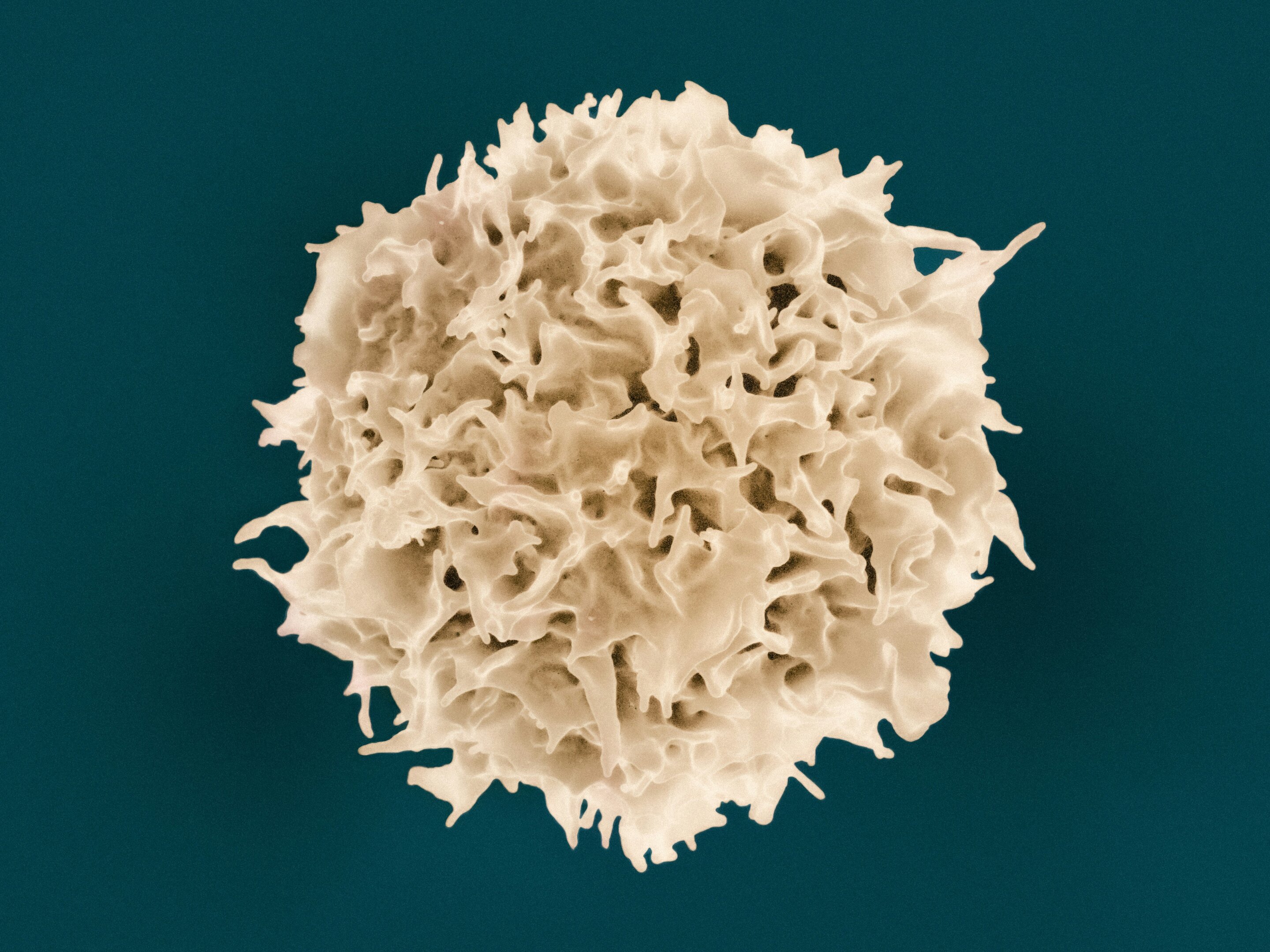
Scientists studying how T cells sense cancer have identified nanoscale contact sites where immune attack signals and braking signals are weighed within seconds. The University of Oxford-led team says the work could help refine checkpoint-blocking cancer immunotherapies.
The study, published in Science Immunology, was conducted by Oxford researchers with collaborators in Austria and China. Lead author Dr. Edward Jenkins and colleagues focused on the earliest moments after a T cell touches a target cell, when the immune cell must decide whether to respond.
T cells are powerful immune cells, and the signals that activate them must be tightly controlled to avoid damage to healthy tissue. Cancer cells can exploit immune checkpoint proteins, including PD-1, to suppress T cell activity and escape immune attack, according to the study.
How do T cells sense cancer cells?
T cells probe other cells using small finger-like projections called microvilli. The Oxford-led study reports that these projections create close nanoscale contacts where T cell receptors and the inhibitory checkpoint protein PD-1 can both gather and send signals.
Jenkins wrote in the paper that lymphocyte activation depends on combining input from several receptors at cell-to-cell contacts. The team found that PD-1 and T cell receptor signals are integrated at microvillar contacts that form during interactions between T cells and their targets.
Using in vitro microscopy, the researchers tested how changes in receptor abundance, receptor domain length, binding strength and cell shape affected signaling. They found that PD-1 inhibitory signaling starts as the close contacts form and can trap the T cell receptor at those sites.
In an accompanying commentary, Dr. Leoma Bere said fluorescence imaging showed PD-1 becoming trapped at microvillar contacts at the interface between cells as those contacts appeared. Once PD-1 was active, the study says it limited the formation of further close contacts and reduced T cell spreading against the target, a step needed for cytotoxic activity.
The researchers also reported that activated PD-1 recruited the signaling molecule SHP2, shortening the duration of T cell receptor signaling. That finding helps explain how an inhibitory checkpoint can quickly restrain an immune response at the exact point where a T cell meets another cell.
What did the study find about nivolumab?
The study also examined nivolumab, a PD-1 checkpoint inhibitor sold as Opdivo and used against many cancers. Under some conditions, the Oxford-led team found that nivolumab briefly encouraged the same type of inhibitory signaling it is meant to stop.
The researchers then designed altered checkpoint-blocking antibodies that did not trap PD-1 at close contacts. In laboratory tests, those modified antibodies produced higher T cell activation, according to the study.
Oxford scientists said the finding could inform the design of next-generation checkpoint therapies. Bere wrote in her commentary that the results may guide antibody design so drugs avoid PD-1 trapping and improve immune checkpoint blockade.
This story draws on original reporting from Medical Xpress.