
REM sleep energy study finds neurons run low during dreaming
A mouse study found brain blood supply rises before REM sleep, while neuronal ATP falls after dreaming sleep begins.
By Tom Brennan · Health & Medicine Correspondent
3 min read
A new REM sleep energy study in mice found that the brain appears to boost its fuel supply around dreaming sleep, yet neurons show a drop in ATP, the molecule they use directly for energy. Tohoku University researchers said the finding adds a metabolic twist to REM sleep, a stage already known for wake-like brain activity while the body remains largely still.
The study, published in Communications Biology, examined how blood flow-related signals and energy molecules change across natural sleep states. The team focused on REM sleep because it is associated with dreaming and memory processing, according to Tohoku University.
Professor Ko Matsui of Tohoku University said the group wanted to examine why vivid dreaming can feel tiring, given that the sleeping brain can be highly active. The researchers described the result as a paradox: more apparent energy supply to the brain during REM sleep did not translate into higher neuronal ATP.
What is the REM sleep energy paradox?
The REM sleep energy paradox is the finding that brain blood volume and astrocytic pyruvate rose around REM sleep, while neuronal ATP fell after REM sleep began, according to the Tohoku University study. ATP is the energy-carrying molecule neurons use for work such as electrical signaling and cellular maintenance.
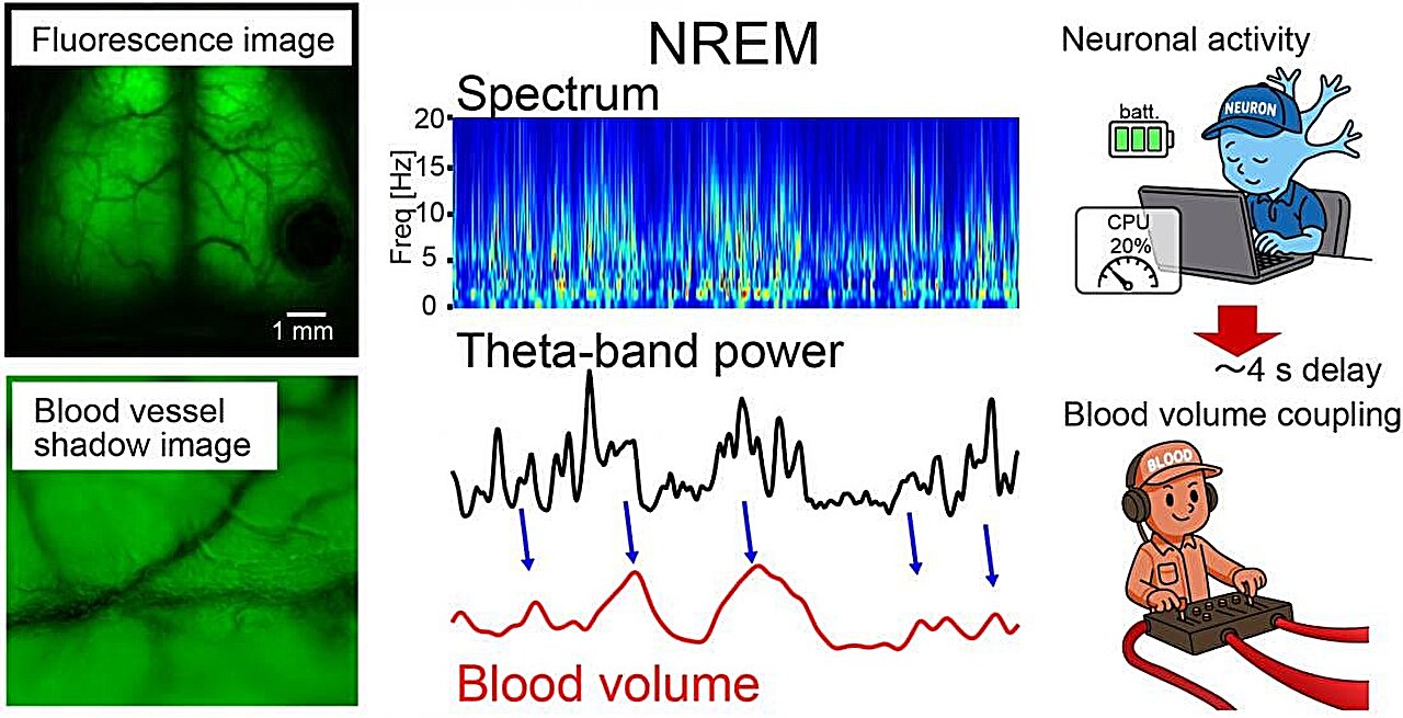
To observe the mouse brain during normal sleep, the team used a UV-curable resin to keep the skull transparent, Tohoku University said. Wide-field fluorescence imaging allowed the researchers to track brain blood volume fluctuations as a sign of fuel supply, neuronal ATP as a direct energy measure in neurons, and astrocytic pyruvate as a molecule tied to glucose-based brain metabolism.
During non-REM sleep, the researchers reported that theta-band brain activity predicted local blood volume changes several seconds later. Tohoku University said that pattern suggests the sleeping brain can adjust blood-vessel responses to match ongoing neural activity and metabolic demand.
The shift into REM sleep looked different. According to the study, brain blood volume began to increase about 50 seconds before REM sleep was identified by electrocorticography, starting in the posterior cortex and spreading toward the front of the cortex.
After REM sleep began, astrocytic pyruvate increased as well, which the researchers said was consistent with greater substrate availability or higher glycolytic activity in astrocytes. At the same time, neuronal ATP decreased, despite the rise in blood volume.
Why might neuronal ATP fall during REM sleep?
The researchers said several explanations are possible. Neurons may be spending large amounts of ATP during REM sleep on processes linked to memory-related synaptic reorganization, communication between the hippocampus and cortex, or broad circuit transitions.
Tohoku University also said energy transfer from astrocytes to neurons may change during REM sleep, or mitochondrial ATP production may shift. The study did not present one settled mechanism for the ATP decline.
Lead investigator Yusuke Takahashi said understanding how the brain balances supply and use may help explain the efficiency of biological intelligence. He said REM sleep offers a natural case in which the brain reorganizes its energy economy during complex internal processing.
The researchers framed the work as part of a broader question in neuroscience: how the brain allocates limited energy across different internal states. Tohoku University said sleep offers a useful way to study that question because the brain remains active even when the body is at rest.
This story draws on original reporting from Medical Xpress.