
New eye imaging method targets noise before scans are built
Researchers describe STOC-T, an OCT-based approach designed to reduce optical crosstalk and sharpen images of retinal cells and other eye structures.
By Priya Raghavan · Science Reporter
4 min read
A new eye-imaging approach aims to make optical coherence tomography clearer by suppressing stray light before it blurs the scan. The method could help researchers see fine retinal structures and cell activity that conventional imaging can miss, according to a paper in the Journal of Biomedical Optics.
Maciej Wojtkowski of the International Centre for Translational Eye Research at the Institute of Physical Chemistry, Polish Academy of Sciences, describes spatio-temporal optical coherence imaging and tomography, known as STOC and STOC-T. The Polish Academy of Sciences said the work focuses on a persistent problem in ophthalmic imaging: useful light returning from tissue can be mixed with scattered light that carries little diagnostic value.
Optical coherence tomography, or OCT, is widely used in ophthalmology to view the retina in cross-section without touching the eye. The Polish Academy of Sciences said clinicians use OCT to detect conditions including glaucoma, age-related macular degeneration, diabetic retinopathy and macular edema before patients may notice vision loss.
How the method addresses optical crosstalk
The problem, as described by Wojtkowski in the journal paper, is optical crosstalk. In scattering tissue, light from one location can reach multiple detector pixels, weakening the match between the image and the structure being examined.
Wojtkowski said the goal in living-tissue imaging is to distinguish light that contains meaningful information from light that degrades the picture, according to the Polish Academy of Sciences. The issue is especially difficult in the retina and choroid, where clinically important structures can measure only a few micrometers.
STOC-T changes acquisition rather than applying a cleanup step after the scan has been recorded, according to the report. The system changes the phase of light entering the tissue using spatial phase masks, then compares and averages the resulting signals.
Scattered light varies across the masks and tends to cancel during averaging, while light tied to stable tissue structure remains, the researchers report. Wojtkowski said the approach is intended to prevent interfering signals from contaminating the image at the measurement stage rather than correcting them afterward.
Tests in tissue models and living eyes
The paper reports laboratory tests using a resolution target covered with either a strongly scattering artificial layer or a 100-micrometer layer of rat skin. According to the Polish Academy of Sciences, the target was hard to see without STOC-T, while phase modulation restored visibility of the structures.
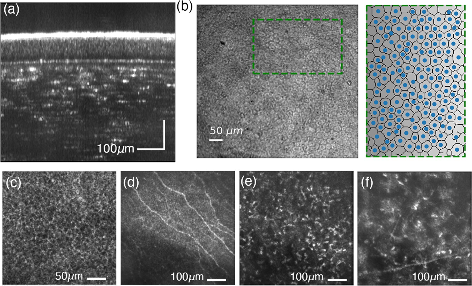
The paper also reports use of STOC-T for retinal imaging. The method visualized retinal layers, photoreceptors, ganglion cells and choroidal microstructure at about 5 micrometers of lateral resolution, approaching cellular scale, according to the researchers.
Wojtkowski also described use of the method for optoretinography, which measures photoreceptor responses to light. The paper reports cone responses to flickering light between 1.5 and 45 Hz, with time constants of about 398 milliseconds and 43 milliseconds, values the researchers said were close to patch-clamp measurements in primate retinas.
The Polish Academy of Sciences said that kind of functional readout may be useful in diseases where cells begin to fail before visible anatomical changes appear. The report did not describe STOC-T as a clinical product.
Technical barriers remain
The World Health Organization says at least 2.2 billion people worldwide have visual impairment, and more than 1 billion cases could have been prevented or remain treatable with earlier and more accurate diagnosis. The Polish Academy of Sciences framed sharper imaging as one route toward earlier detection and monitoring.
The journal paper lists substantial engineering hurdles. STOC-T requires a high-speed CMOS camera of 512 by 512 pixels running at 60,000 frames per second, a tunable laser in the 800-to-870 nanometer range and heavy data handling, with a single acquisition exceeding 8.5 GB.
The researchers also discuss multimode optical fibers as a possible way to produce phase modulation. A fiber with a 50-micrometer core and 300-meter length could support about 800 propagation modes and, in theory, reduce optical crosstalk noise nearly 29-fold without active control electronics, according to the paper.
Wojtkowski said the next technical targets include speed, data volume, phase encoding and automated reconstruction, according to the Polish Academy of Sciences. The study, “Spatio-temporal optical coherence imaging and tomography for in vivo applications,” was published in the Journal of Biomedical Optics.
This story draws on original reporting from Medical Xpress.