
Neurosurgeon eye tracking AI research maps expert scan reading
Macquarie University studies used fractal math to measure how medical image expertise develops and how AI might learn from expert gaze.
By Tom Brennan · Health & Medicine Correspondent
3 min read
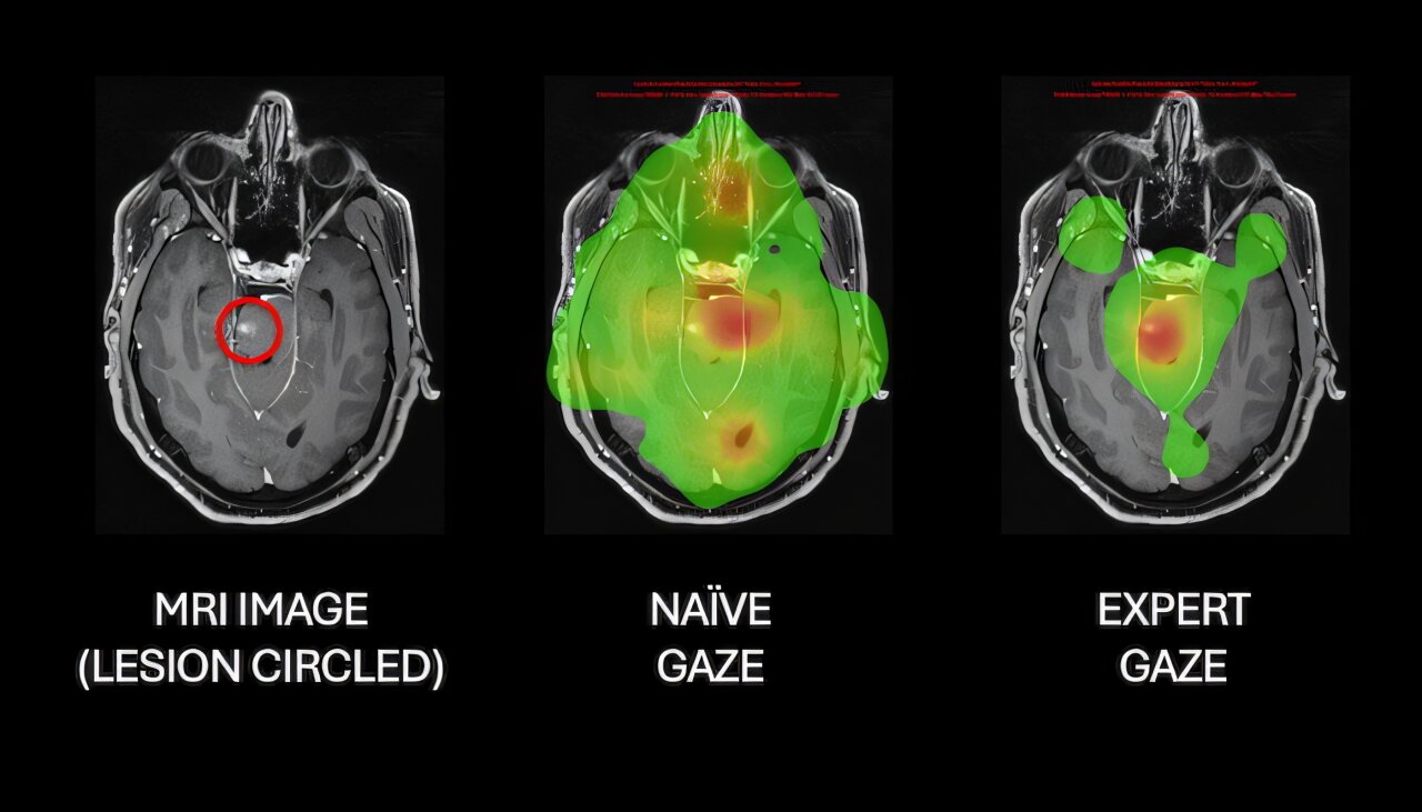
Neurosurgeon eye tracking AI research from Macquarie University suggests expert scan reading leaves measurable patterns in the way doctors look at medical images. The work matters because those patterns could help assess medical trainees and shape future systems that interpret complex scans.
Researchers in Macquarie University’s Computational NeuroSurgery Lab, led by Professor Antonio Di Ieva, reported the findings in two 2026 studies. The team used high-resolution eye tracking, fractal mathematics and machine learning to compare how novices, trainees and qualified neurosurgeons inspect X-rays, CT images and MRI scans.
The studies build on the lab’s view that medical image expertise is reflected not only in what a doctor knows, but also in how the eyes search for relevant information. The researchers converted gaze patterns into numerical measures so they could compare the structure and complexity of visual scanning across experience levels.
How can eye tracking help train AI?
Eye tracking records where a viewer looks and for how long, while fractal analysis measures how organized or complex the overall gaze pattern is. In this work, Macquarie researchers used those measurements as a possible signal of expert visual judgment that machine-learning systems could learn from.
Dr. Ghasem Azemi, a research associate in the lab, said fractal analysis can show whether gaze is scattered or follows more ordered patterns. That approach goes beyond timing individual fixations and gives researchers a way to quantify the development of expertise, according to Macquarie University.
In one study, published in Medical & Biological Engineering & Computing, researchers followed 13 Doctor of Medicine students from Macquarie Medical School over three consecutive semesters. The students viewed a range of medical images while their eye movements were recorded.
The team combined the gaze measurements into what it called the Fractal Eye-Gaze Expertise Index. According to the study, the index showed a statistically significant trend as students advanced: their viewing patterns became more organized and closer to expert scan-reading behavior.
Azemi said the shift suggested students were moving away from a broad response to basic image features and toward more targeted visual processing tied to diagnosis. The researchers said fractal metrics made that change measurable during training.
What did the brain MRI study find?
A second study, published in the Journal of Eye Movement Research, examined 69 participants. The group included naïve observers, neurosurgery registrars and qualified neurosurgeons who viewed normal and abnormal brain MRI scans.
Macquarie researchers found that experts inspected the images more efficiently, looked longer at diseased areas and handled image complexity differently depending on the type of pathology. The study also tested an AI model that combined fixation duration with 3D fractal dimension.
That model separated naïve, trainee and expert viewers with accuracy above 93%, according to the study. The result suggests gaze behavior can carry enough information to help classify levels of visual expertise in brain MRI interpretation.
Di Ieva, who founded the Computational NeuroSurgery Lab in 2018, said the research began with a question about how expert cognition might be transferred into computer vision. He said the findings point to possible uses in medical trainee assessment, radiology and neurosurgery curriculum design, and AI systems built to interpret medical images in ways closer to expert clinicians.
This story draws on original reporting from Medical Xpress.