
MIT team improves portable 3D ultrasound for breast imaging
Researchers say the system could support more frequent breast checks and long-term monitoring without a trained ultrasound operator.
By Tom Brennan · Health & Medicine Correspondent
3 min read
MIT researchers have reported upgrades to a portable 3D breast ultrasound system that they say improve image quality and make the device easier for non-specialists to use. The work matters because mammograms performed once a year can miss cancers that arise between screenings, particularly for people at higher risk.
The study, published in Nature Communications, describes a compact ultrasound approach intended for repeated breast imaging in a clinic or potentially at home. According to MIT, the system is designed to help detect possible tumors, cysts and microcalcifications, and to support monitoring after breast cancer treatment.
Canan Dagdeviren, an associate professor at MIT’s Media Lab and senior author of the study, has focused on more frequent screening after an aunt died of interval breast cancer in 2015, MIT said. The researchers said interval cancers account for 20% to 30% of breast cancer cases and tend to be more aggressive.
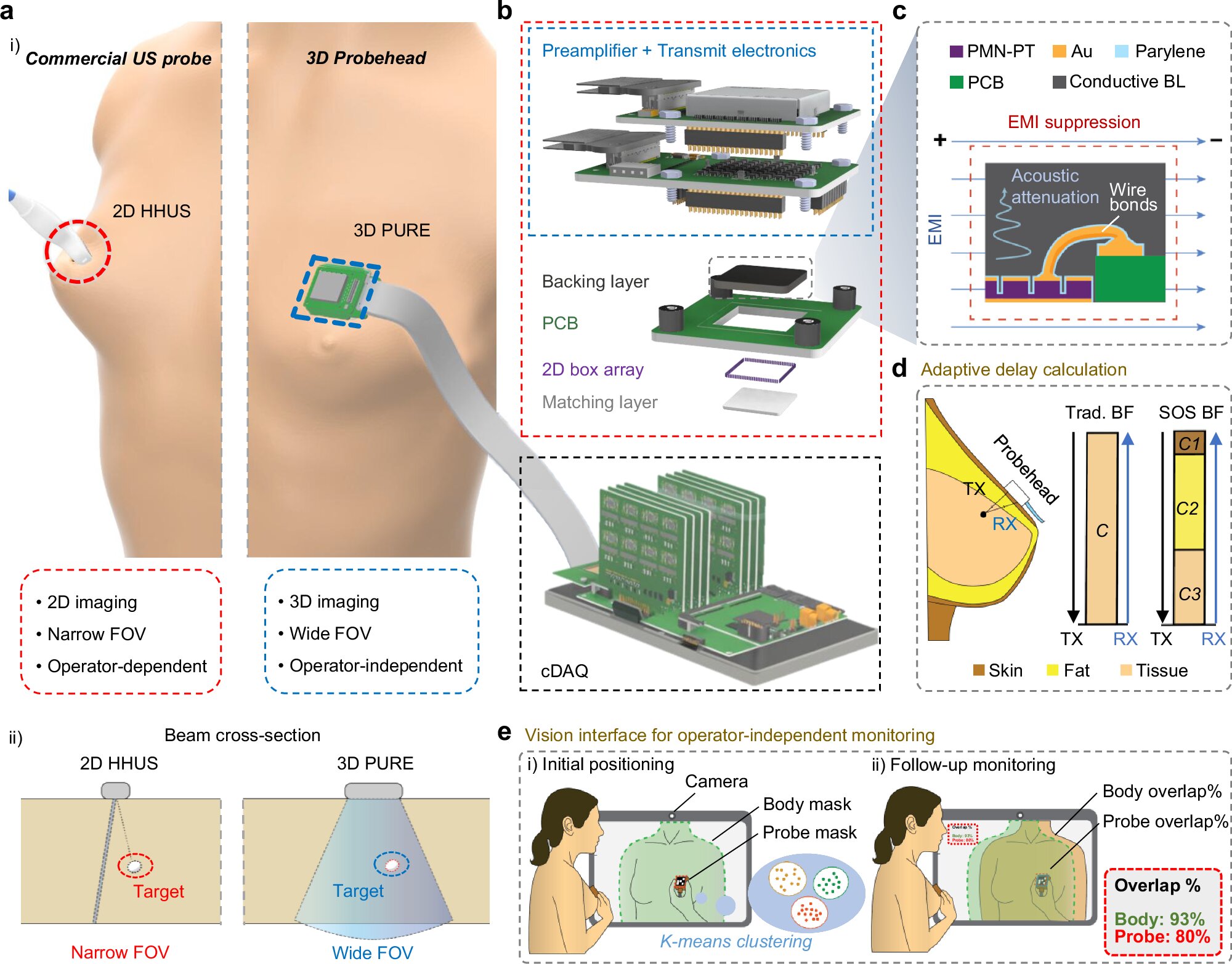
Sharper images from a portable probe
MIT said the team previously demonstrated a small ultrasound probe linked to an acquisition and processing module slightly larger than a smartphone. That earlier version could produce a 3D image of the whole breast by scanning two or three positions, according to MIT.
In the new study, the researchers added a backing layer to the ultrasound transducer. According to the paper, that layer helps control the sound waves, improves image resolution, broadens the operating frequency range and reduces acoustic and electrical noise.
The team also developed an image-processing algorithm that adjusts beamforming based on estimated differences in how sound travels through tissue. MIT said the method is intended to account for tissue types such as skin and fat, and graduate student Shrihari Viswanath said the approach produced up to a 10% resolution improvement.
To test usability for finding small targets, the researchers asked 10 volunteers without ultrasound expertise to scan a tissue phantom, a gel-like material made to mimic human tissue. MIT said participants located embedded microtargets at a higher rate with the new system than with a conventional ultrasound probe.
Interface aims to reduce operator dependence
The researchers also built a computer-screen interface that guides users to place the probe in the right position, according to MIT. The team said repeatable placement could be useful for tracking tissue over time, including during neoadjuvant therapy or when monitoring known findings such as fibroadenomas or microcalcifications.
In a separate trial involving seven people, MIT said users were able to put the probe in the correct spot each time they scanned. Graduate student Hyeokjun Yoon said the interface uses computer vision and shows live images, reducing the need for a trained operator to move the probe around the breast.
The study’s lead authors are former MIT postdoctoral researcher Md Osman Goni Nayeem and MIT graduate students Viswanath and Yoon. The paper is titled “Portable, real-time 3D ultrasound for operator-independent breast imaging,” according to Nature Communications.
MIT said future versions may use a phone or tablet interface to make the system more portable. Dagdeviren and some students also hope to form a company to pursue commercialization, with breast cancer diagnosis as the first target and other soft-tissue uses, including ovarian cancer, endometriosis monitoring and fetal monitoring, as possible later applications.
This story draws on original reporting from Medical Xpress.