
Light microscopy study aims to sharpen thyroid cancer diagnosis
Houston Methodist researchers say SHG microscopy can read collagen changes in thyroid tissue, a step they hope will improve diagnosis and limit unnecessary surgery.
By Tom Brennan · Health & Medicine Correspondent
2 min read
Houston Methodist researchers report that a light-based microscopy method may help doctors identify papillary thyroid cancer by measuring collagen changes in thyroid tissue. If confirmed in larger groups, the team says the approach could add more objective information to current testing and may help some patients avoid surgery.
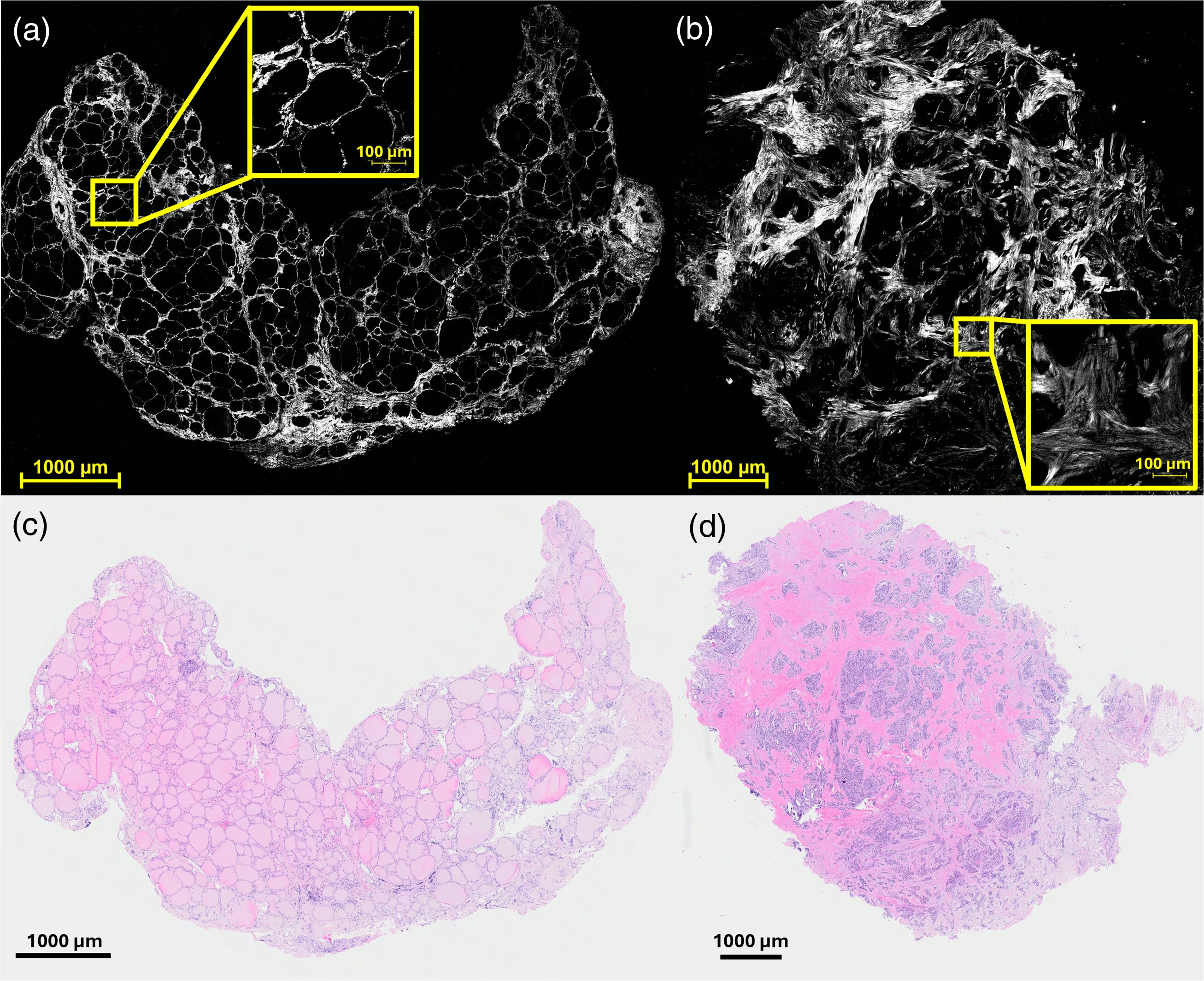
The study, published in the Journal of Biomedical Optics, focused on second harmonic generation microscopy, or SHG. Houston Methodist said the technique uses light to study collagen, a structural protein in tissue, and can reveal patterns that may differ between cancerous and noncancerous thyroid samples.
Papillary thyroid cancer is the most common form of thyroid cancer, according to Houston Methodist. The National Institutes of Health says thyroid cancer is the most common cancer of the endocrine system and the leading cancer among people ages 16 to 33.
How the method works
According to the researchers, SHG microscopy examines collagen without relying on the same kind of subjective visual assessment used in some conventional pathology workflows. The Houston Methodist team said changes in collagen organization may act as measurable signs that a thyroid nodule is malignant.
Stephen Wong, the John S. Dunn Presidential Distinguished Chair in Biomedical Engineering at Houston Methodist, co-led the work with Raksha Raghuanthan, an assistant professor at the Houston Methodist Research Institute. Wong said the approach could make thyroid cancer diagnosis quicker, more accurate and more consistent while supporting, rather than replacing, conventional cytology.
Wong also said the study used interpretable statistical modeling to link collagen patterns with thyroid cancer. He contrasted that approach with artificial intelligence systems whose decision-making can be harder for clinicians to examine.
Potential clinical role
Houston Methodist said the method could become a less invasive addition to standard laboratory testing if further research supports the findings. The researchers said clearer and more consistent tissue information could help doctors make better diagnostic decisions for patients with thyroid nodules.
The team’s goal is to develop the approach as a biopsy-related method and cut down on invasive surgeries, according to Houston Methodist. The next steps include testing the method in larger patient cohorts and assessing whether the technology can distinguish other thyroid cancer subtypes.
The study’s publication lists Wesley Poon and colleagues as authors under the title “Quantitative second harmonic generation microscopy for characterizing collagen remodeling in papillary thyroid carcinoma.” The paper appeared in the Journal of Biomedical Optics in 2026.
Collaborators included Lin Wang, Hong Zhao, Helmi Khadra and Elizabeth Jacobi of Houston Methodist; Wesley Poon, Orhun Davarci and Reid Master of Texas A&M University; and Jun Liu of Shanghai Jiao Tong University, according to Houston Methodist.
This story draws on original reporting from Medical Xpress.