
Human red blood cell development differs from mouse model, study finds
A Nature Genetics study reports that human red blood cell clusters form without the macrophage hub long seen in mice.
By Tom Brennan · Health & Medicine Correspondent
3 min read
Human red blood cells appear to mature in a structure different from the one long inferred from mouse research, according to a Northwestern Medicine-led study published in Nature Genetics. The finding matters because mouse models guide much of the research into blood disorders and possible treatments.
The team studied erythroblastic islands, microscopic sites where developing red blood cells mature. Peng Ji, senior author of the study and a professor of pathology at Northwestern University Feinberg School of Medicine, said decades of understanding about these sites have relied heavily on mouse experiments and cell studies that remove tissue from its normal setting.
To examine the structures in place, Ji and colleagues used spatial transcriptomics, a method that maps gene activity across intact tissue. Northwestern Medicine said the approach allowed the researchers to compare mouse and human samples while preserving the organization of the cells.
Different architecture in humans
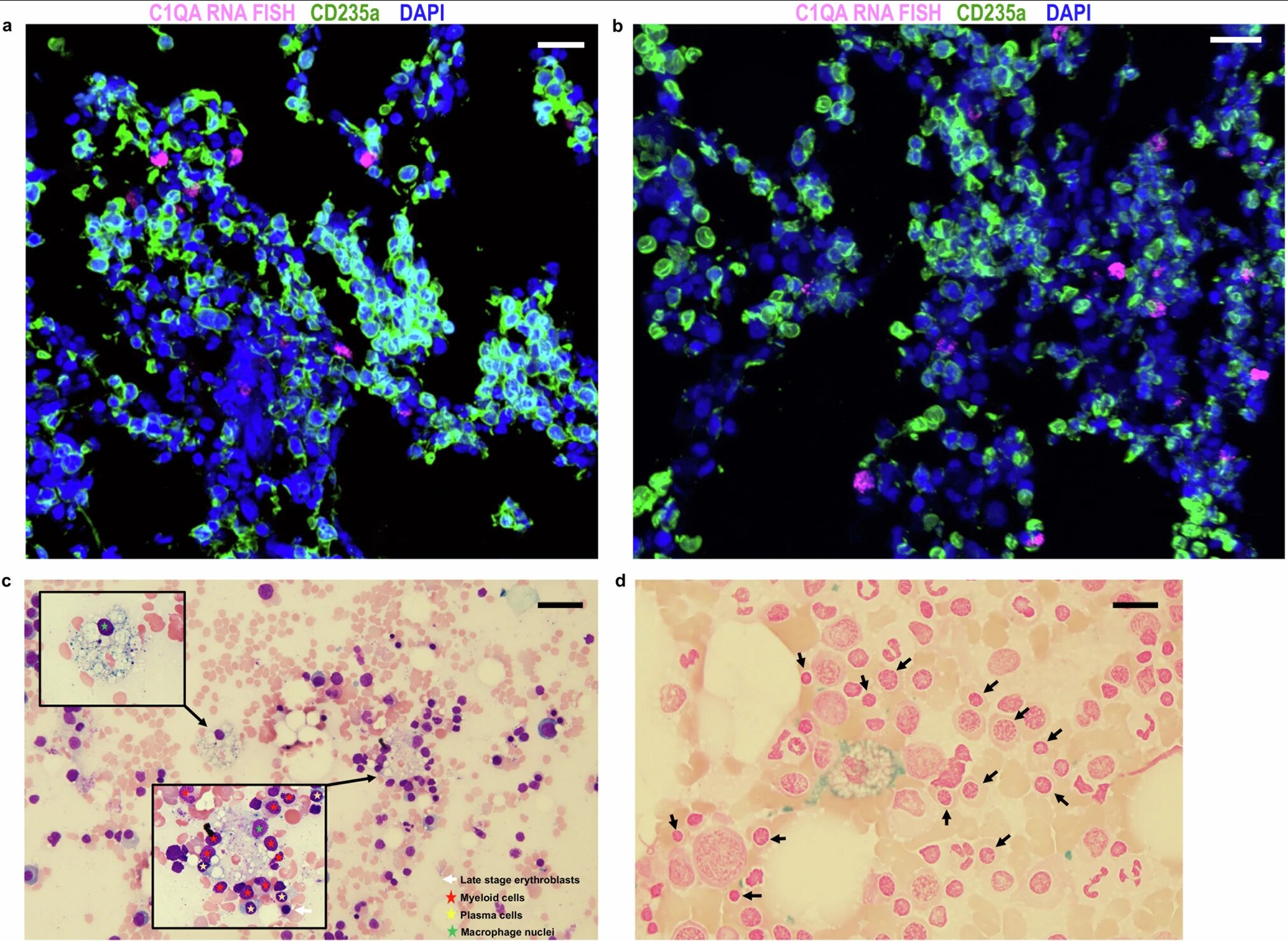
The study reports that mouse erythroblastic islands fit the conventional model: developing red blood cells cluster around a macrophage, a type of white blood cell. In mice, the researchers found a macrophage marked by the protein C1q at the center of the cluster, where it helps clear cellular material during red blood cell maturation.
Human samples did not show the same arrangement, according to the study. Instead of organizing around a central macrophage, human erythroid cells formed clusters on their own, with the molecule ICAM4 helping the cells attach to one another.
Ji said the species-specific structure was the study’s most unexpected result. He said the work challenges the assumption that human blood formation matches the pattern observed in mice.
The researchers described the finding as a major shift in understanding how the body produces red blood cells, which Ji called the body’s most abundant cell type. He said differences between mouse and human biology can affect how scientists interpret disease mechanisms and design therapies.
Possible link to blood disease
The team also examined bone marrow samples from patients with myelodysplastic syndromes, or MDS, a blood disease that often appears as anemia. Northwestern Medicine said the human erythroblastic island structures were disrupted in those samples but could be partly restored after treatment.
The study suggests that the organization of these cell clusters may be tied to disease progression and recovery. Ji, who is also a member of the Robert H. Lurie Comprehensive Cancer Center of Northwestern University, said a more accurate human model gives researchers a better basis for studying blood diseases.
The findings also leave an open question about cellular cleanup in humans. In mice, macrophages help remove nuclei that are expelled as red blood cells mature, according to the study.
Ji said future work will test whether other scavenger cells or different mechanisms perform that role in humans. He said the broader goal is to develop models and therapies that better reflect how human systems work.
The paper, titled “Spatial transcriptomic analyses highlight distinct erythroid niches in mice and humans,” was authored by Xu Han and colleagues and published in Nature Genetics.
This story draws on original reporting from Medical Xpress.