
Core2Edge glioblastoma model maps tumor spread in human brain tissue
Bonn researchers describe a human-tissue model that tracks glioblastoma invasion down to single cells, aiming to study why tumors recur.
By Priya Raghavan · Science Reporter
3 min read
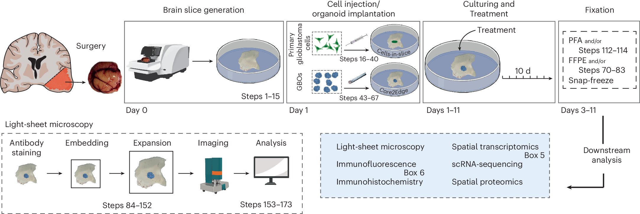
The Core2Edge glioblastoma model lets researchers follow aggressive brain tumor cells as they move from a tumor mass into human brain tissue, according to University Hospital of Bonn. The method, described in Nature Protocols, matters because glioblastoma cells that spread beyond the visible tumor can remain after treatment and contribute to recurrence.
University Hospital of Bonn said glioblastoma is among the most aggressive human cancers and remains incurable despite surgery, radiation and chemotherapy. The hospital said one major challenge is invasion: tumor cells can migrate into apparently healthy brain regions, making complete surgical removal impossible.
What is the Core2Edge glioblastoma model?
Core2Edge is a laboratory system that combines patient-derived glioblastoma organoids with living slices of human brain tissue, according to the Bonn team. The goal is to reproduce the path from the tumor core to scattered individual cancer cells at the edge of invasion.
The researchers used tumor organoids grown from freshly removed patient material, University Hospital of Bonn said. They placed those organoids into organotypic human brain slice cultures made from tissue removed during neurosurgical access procedures that would otherwise have been discarded.
Dr. Matthias Schneider, deputy director of the Department of Neurosurgery at University Hospital of Bonn and head of the Brain Tumor Translational Research Group at the hospital and the University of Bonn, said the model is designed to study tumor cells that remain hidden in the brain after surgery. According to the hospital, Schneider said Core2Edge is based entirely on human tissue and closely reflects features seen in patients.
How the researchers tracked invading cells
The Bonn team used high-resolution light-sheet fluorescence microscopy to image tumor spread, according to the Nature Protocols paper. After the tissue was fixed, the researchers expanded it evenly to improve light penetration and make fine structures easier to see.
The sample was then scanned in layers to build three-dimensional views of tumor-infiltrated brain tissue, University Hospital of Bonn said. Ahmad Melhem, first author of the study and co-developer of the model during his doctoral research, said the approach allowed the team to measure tumor spread in 3D and examine the shape of individual invading cells.
The researchers also used spatial transcriptomics, a method that shows which genes are active in cells and where those cells sit in tissue, according to the hospital. The team said this is relevant in glioblastoma because cells within one tumor can show different patterns of gene activity, a feature known as intratumoral heterogeneity.
University Hospital of Bonn said that heterogeneity is considered a driver of treatment resistance because some cell populations can survive radiation and chemotherapy more effectively and later fuel renewed tumor growth. Dr. Anna-Laura Potthoff, a neurosurgeon and clinician-scientist in the Brain Tumor Translational Research Group, said Core2Edge reproduced intratumoral heterogeneity, supporting the view that the model reflects patient tumors.
The Bonn researchers said the model could support future work on which cellular programs operate in invasion zones and which treatment targets may help delay or prevent recurrence. Schneider also said Core2Edge could reduce reliance on animal experiments by allowing key aspects of glioblastoma biology, including invasion and heterogeneity, to be studied directly in human tissue.
This story draws on original reporting from Medical Xpress.