
Brain metabolism may help predict lung cancer survival, study finds
A Journal of Nuclear Medicine study links lower brain FDG uptake to higher mortality in advanced non-small cell lung cancer.
By Tom Brennan · Health & Medicine Correspondent
3 min read
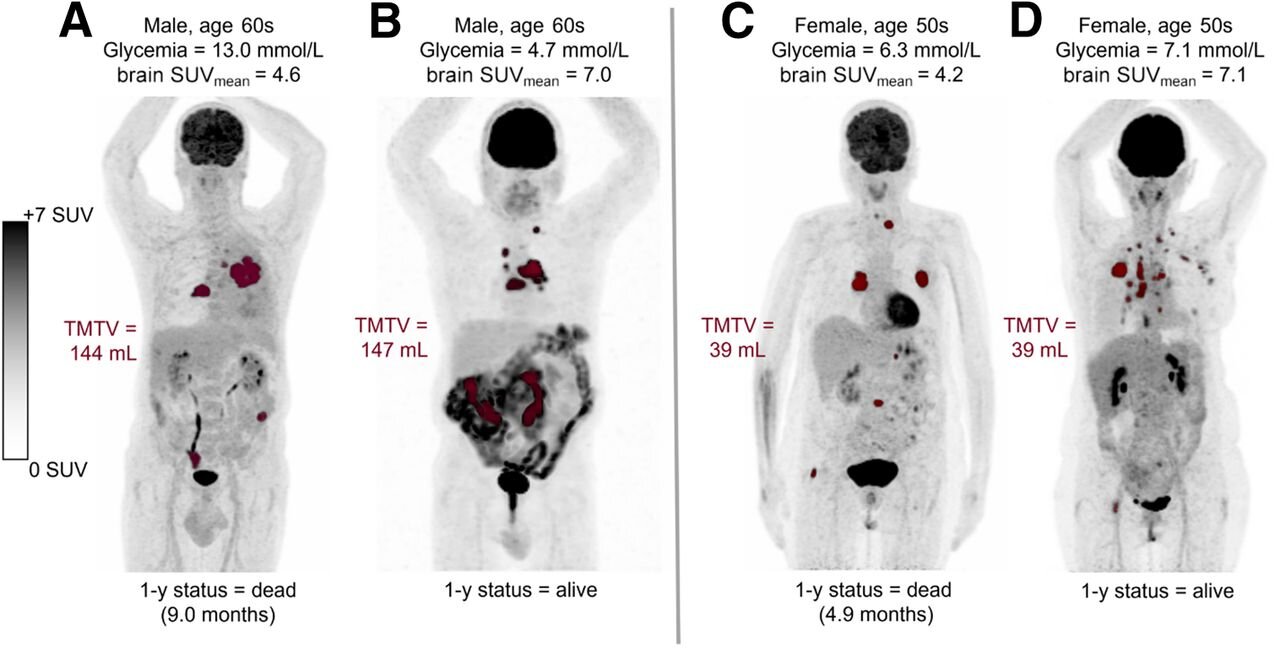
Brain metabolism lung cancer survival signals may be available from scans patients already receive, according to research published in The Journal of Nuclear Medicine. The study found that lower brain metabolic activity on 18F-FDG PET/CT was tied to higher mortality in people with advanced non-small cell lung cancer.
The findings matter because metastatic lung cancer can be hard to treat and patients vary widely in how long they benefit from therapy. The Society of Nuclear Medicine and Molecular Imaging said adding brain 18F-FDG uptake to clinical, tumor and biological information improved predictions of overall survival, which could help doctors identify patients who may need earlier supportive care.
Non-small cell lung cancer, or NSCLC, is the most common broad category of lung cancer. The research focused on advanced disease, where treatment decisions are often difficult because some patients resist therapy or see only brief benefit without a meaningful survival gain, according to the society.
How does brain metabolism predict lung cancer survival?
The researchers found that patients with higher brain metabolism tended to live longer overall. Brain metabolism was significantly lower among patients who died within one year than among those who were alive at that point, according to the study.
Brain metabolic activity also stood out as an independent prognostic factor for overall survival. The study reported that it was linked with several clinical and blood biomarkers while still adding separate information, meaning it was not just repeating what those measures already showed.
18F-FDG PET/CT is an imaging method that uses a radioactive glucose tracer to show how tissues use energy. Because many cancer patients already undergo this type of scan as part of routine care, the brain metabolism measurement could be drawn from existing imaging rather than requiring another test, Julie Auriac, Ph.D., said in a statement released by the society.
Auriac, a research engineer at the Imaging, Innovative Radiotherapy, and Systems Medicine Laboratory in Orsay, France, said recent work has suggested that metabolic activity and tissue density measured on whole-body 18F-FDG PET/CT can help predict lung cancer outcomes by reflecting a patient’s overall condition. She said the study was designed to examine whether brain metabolic activity measured on those scans was associated with survival in advanced NSCLC.
What did the study examine?
The retrospective study included patients with advanced NSCLC who had pretreatment 18F-FDG PET/CT scans between 2010 and 2023. Researchers gathered clinical and biological data, tumor characteristics and brain metabolism measures, then assessed how well those features predicted overall survival.
The team also examined how brain metabolism related to clinical, imaging and blood biomarkers. According to the published findings, brain metabolism remained useful alongside those other measures rather than being fully explained by them.
Lung cancer remains a leading cause of cancer deaths worldwide, according to the society. It said average overall survival ranges from more than three years in early-stage disease to less than five months in metastatic disease, despite advances in treatment.
Auriac said the results support viewing cancer as a systemic disease. She said prognosis may depend not only on tumor features, but also on information from other organs and tissues that has not been widely used in outcome prediction.
This story draws on original reporting from Medical Xpress.