
AI ultrasound imaging work at UVA targets clearer sonograms
UVA Ph.D. candidate Soumee Guha is building diffusion models to reduce distortion in ultrasound and other medical images.
By Tom Brennan · Health & Medicine Correspondent
3 min read
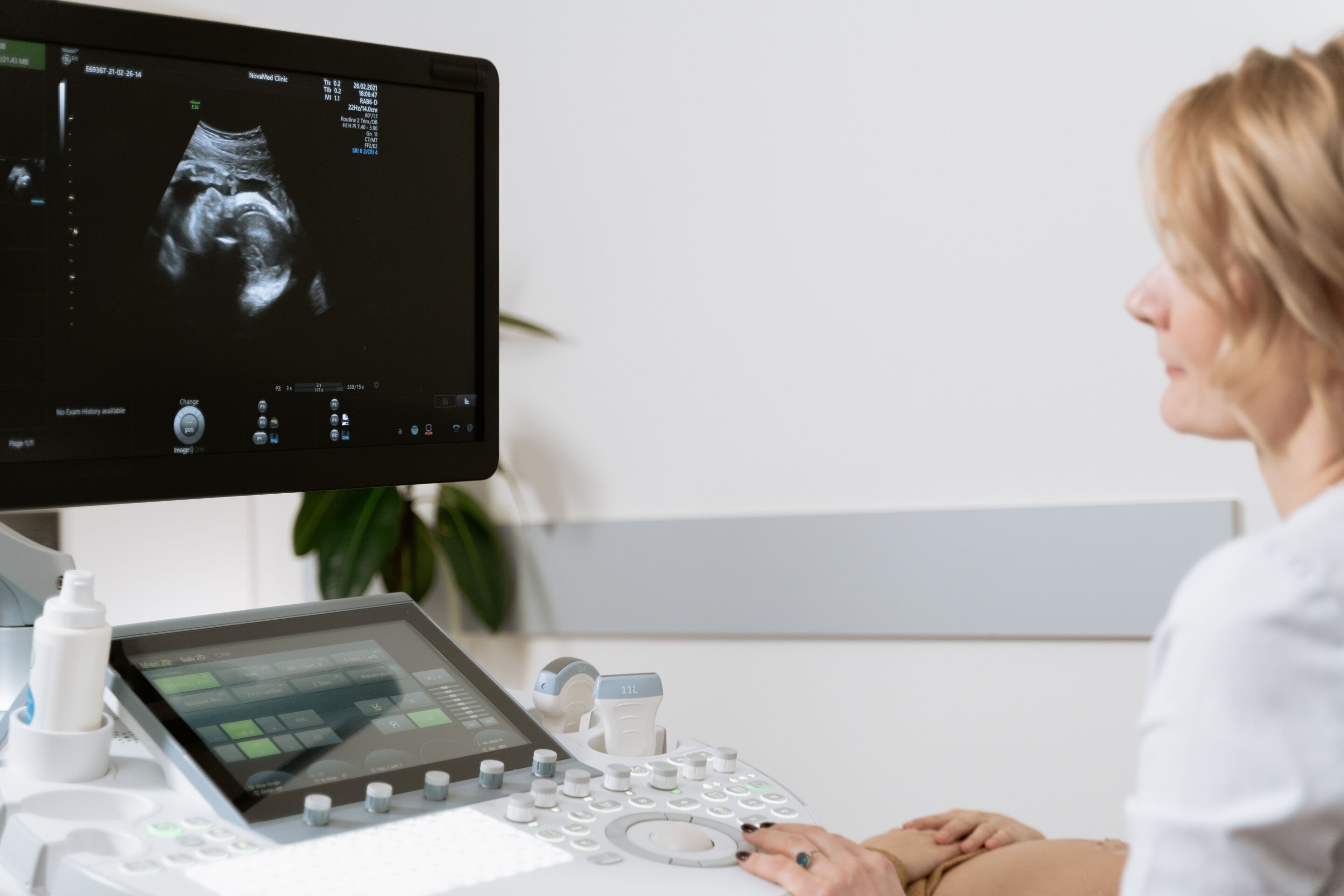
A University of Virginia doctoral student is developing AI ultrasound imaging methods that could make sonograms and other medical scans easier for clinicians to read. The work matters because ultrasound images can be grainy or distorted, which UVA says may limit their diagnostic value in pregnancy care and other medical uses.
Soumee Guha, a Ph.D. candidate in UVA’s Department of Electrical and Computer Engineering, builds generative artificial intelligence models for medical imaging, according to the university. UVA said her approach is designed for areas including fetal imaging, liver disease assessment and heart-function analysis.
The university said Guha’s research differs from many diagnostic AI projects because it does not depend on large stores of patient images for training. Guha uses mathematical tools and information about how imaging systems work to guide the models, according to UVA.
How can AI improve ultrasound imaging?
Ultrasound uses a transducer to send sound waves through the body and record the signals that return from different depths and angles, UVA said. That process can produce distorted images, including the familiar speckled look of sonograms.
Speckling is a granular pattern caused when sound or light waves scatter from points on an imaged surface, Guha said in UVA’s account of the work. Similar distortion can affect endoscopic images collected inside the gastrointestinal or respiratory tract when doctors look for signs of disease.
Guha’s models are diffusion models, a form of generative AI. According to UVA, her system starts with distorted images, spreads visual noise through the image field and then reduces that noise while using learned patterns and mathematical constraints to produce a clearer version.
The result, Guha said, can give clinicians two image sets to evaluate: the original scan from the medical device and an AI-generated version. Doctors could compare them, use them together or examine differences between what the system captured and what the model expected, according to UVA.
Why Guha’s method uses physics
Many machine-learning image tools need large training sets, which can be expensive and hard to obtain in medicine, UVA said. Guha’s dissertation work adds mathematical constraints tied to the imaging system and its capture mode, aiming to improve accuracy with less dependence on patient data.
Guha said image-enhancement models that do not account for the physics of the imaging device may perform poorly in clinical or research settings. Her work incorporates established physical models into the algorithmic framework, according to UVA.
Scott Acton, Guha’s adviser and chair of UVA’s electrical and computer engineering department, said Guha is the first researcher to develop diffusion models for de-speckling. He also said she is the first to address a blur problem tied to how imaging systems capture point sources of light.
The de-speckling methods in Guha’s dissertation could apply beyond ultrasound, UVA said. The university said related models may improve sonar, radar and laser images, which can also be degraded by speckling.
What comes next for the research?
Guha’s doctoral project, “Learning Under Multiplicative Noise: Principled Image Enhancement Frameworks for Coherent Imaging,” began in fall 2021, according to UVA. She defended the dissertation this spring and is preparing to graduate.
Acton said Guha’s AI-generated images could be used to train future AI models and evaluate tasks such as tumor classification or volume measurement. UVA said Guha is seeking postdoctoral research opportunities to continue work in biomedical imaging.
This story draws on original reporting from Medical Xpress.