
AI brain tumor MRI system detects scans, then classifies tumor type
University of Sharjah researchers report a two-stage MRI system that detects tumors and sorts them as glioma, meningioma or pituitary tumors.
By Tom Brennan · Health & Medicine Correspondent
3 min read
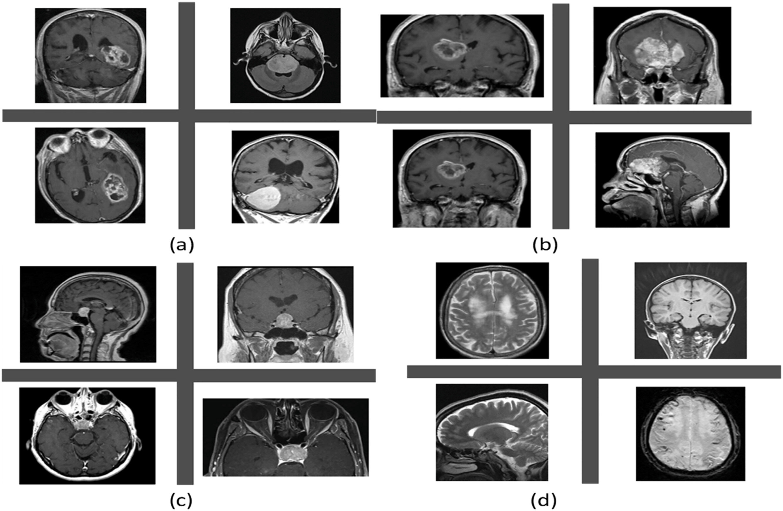
Researchers led by the University of Sharjah have developed an AI brain tumor MRI system designed to first detect whether a scan shows a tumor and then classify the tumor type. The approach matters because faster, more accurate MRI analysis could support earlier screening, though the authors say more clinical validation is needed before routine use.
The findings were reported in Healthcare Analytics. According to the study, the researchers compared several deep learning methods under the same image-processing and testing setup rather than proposing a single new architecture.
What is the AI brain tumor MRI system?
The system uses a two-step workflow for brain MRI images, according to the University of Sharjah-led team. In the first step, an AI model separates tumor scans from nontumor scans; in the second, scans flagged as tumors are sorted into glioma, meningioma or pituitary tumor categories.
Deep learning models learn visual patterns from large sets of images. In this study, the team examined how convolutional neural networks, long short-term memory networks, attention mechanisms and Vision Transformers performed on the same diagnostic tasks.
How the models performed
The researchers reported that performance improved when standard convolutional neural networks were combined with long short-term memory layers and attention mechanisms. The authors said attention helped the models capture broader image patterns that were useful for classification.
For distinguishing among the three tumor types, the study found that a CNN-LSTM model with attention was especially effective. For the first-stage task of separating tumor from nontumor scans, the researchers reported that a Vision Transformer delivered the strongest performance among the models tested.
The authors said those results suggest the models could help build early tumor screening tools for clinical workflows. They did not present the work as ready for deployment, and said additional testing would be needed before use in everyday medical practice.
What data did the study use?
The study analyzed 7,023 MRI images, including glioma, meningioma and pituitary tumor images as well as images from healthy individuals, according to the authors. The researchers said all experiments followed a defined image-level evaluation protocol to improve transparency and reproducibility.
The team applied a common preprocessing process to all images. According to the study, that included resizing scans to a fixed input resolution, normalizing intensity and equalizing contrast to reduce differences among datasets.
The authors also noted a limitation: full patient identifiers were not consistently available across the public datasets. Because of that, the study could not guarantee strict patient-level separation between training and testing data.
How fast is the system?
The researchers reported average inference times of 21.6 milliseconds for the tumor-versus-nontumor task and 17.6 milliseconds for the multiclass tumor-type task. The authors said those speeds support the possibility of near-real-time clinical applications.
The study places the work in the broader context of brain tumors as a global health problem. The World Health Organization estimates brain tumors account for about a quarter of a million deaths each year, according to the researchers.
The authors said future work should test the models on larger and more diverse clinical datasets, including images from different hospitals and equipment. They also suggested adding interpretability tools, such as feature attribution and lesion-level visualization, to help clinicians understand how the AI reaches its decisions.
This story draws on original reporting from Medical Xpress.