
New 3D nerve maps of rat knees may aid research on jaw pain
University of Pittsburgh researchers used tissue clearing and light-sheet microscopy to trace nerves in rat knee joints, a step toward studying TMJ pain.
By Priya Raghavan · Science Reporter
3 min read
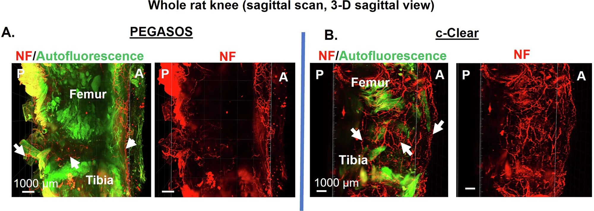
University of Pittsburgh researchers have produced three-dimensional images of nerve networks inside rat knee joints, work they say could help clarify why some jaw joint disorders cause pain. The study gives scientists a way to view nerve branching in dense joint tissue without cutting the tissue into thin sections.
The findings were published in npj Imaging in a paper on tissue clearing and 3D imaging of neural innervation in rat knee joints. Alejandro Almarza, a professor of oral and craniofacial sciences at Pitt’s School of Dental Medicine with a secondary appointment in bioengineering, led the work with collaborators at Pitt’s Center for Biologic Imaging.
Almarza’s target is the temporomandibular joint, or TMJ, which connects the jawbone to the skull on each side of the face. Pitt said temporomandibular disorders include more than 30 conditions involving jaw pain and dysfunction, and the causes of many of those problems remain poorly understood.
Why knees were used
The team used rat knee tissue as a test case because joint tissue is dense and hard to image, according to Pitt. Traditional approaches rely on slicing samples and staining them so nerve cells can be seen under a microscope, but that process removes the full three-dimensional structure of the joint.
To preserve that structure, the researchers combined tissue clearing with light sheet fluorescence microscopy. Pitt said tissue clearing makes a sample transparent enough for imaging, while the microscope scans the tissue volume with a sheet of light, reducing damage and speeding the process compared with conventional microscopy.
The imaging work drew on technology and protocols developed at Pitt’s Center for Biologic Imaging. Simon Watkins, a distinguished professor of cell biology and immunology who founded the center, contributed custom microscope expertise, while Alan Watson, an associate professor of cell biology, contributed clearing methods, computing systems and analysis tools, according to the university.
Two clearing methods compared
The study compared PEGASOS, an established clearing method for tissue containing bone, with c-Clear, a method developed at the Pitt imaging center. According to the researchers, PEGASOS left autofluorescent material that raised background signal and limited laser penetration through the sample.
The c-Clear method added a 24-hour photobleaching step before staining, which Pitt said helped deactivate those molecules. That allowed fluorescent antibodies to bind to neurofilament, a nerve marker, and generated a complete 3D view of nerves running through the joint.
Almarza said the c-Clear workflow takes about six to eight weeks to produce an image, making it more time- and labor-intensive than standard histology. The payoff, he said, is a clearer view of how nerves branch through a joint.
Large files, public data
The images created substantial computing demands. Pitt said one 3D nerve map of a rat knee contains about 1 terabyte of data, while the full project generated about 16 terabytes.
The Center for Biologic Imaging used 7 petabytes of storage and an H200 GPU cluster to stitch, clean and analyze the data sets, according to Pitt. The team deposited the collection on the National Institutes of Health’s SPARC Portal for public access.
The work is tied to the NIH HEAL Initiative through the ReJoin Consortium, which is mapping nerve architecture across joints, species and disease states. Pitt said validating c-Clear on difficult joint tissue now gives Almarza’s group a path toward studying nerve patterns in the TMJ and asking why imaging findings and pain do not always match in people with jaw joint problems.
This story draws on original reporting from Medical Xpress.