
Sst-Chodl neurons promote sleep in mice, study finds
Activating a rare group of cortical neurons increased sleep in mice, challenging the view that the cortex only follows deeper sleep signals.
By Hana Yoshida · Markets Reporter
3 min read
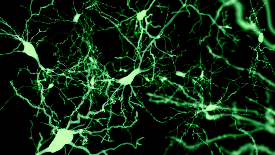
Rare cortical cells called Sst-Chodl neurons promoted sleep when researchers activated them in mice, according to a Nature study reported by Ars Technica. The result suggests the cerebral cortex can help start sleep-related rhythms, rather than only responding to commands from deeper brain regions.
The experiments do not point to a sleep treatment for people. They used optogenetic stimulation, a technique that makes genetically targeted cells fire in response to light, in mice; the cells’ natural triggers and their role in human sleep remain unresolved.
What are Sst-Chodl neurons, and how did they affect sleep in mice?
Sst-Chodl neurons are inhibitory nerve cells in the cerebral cortex, identified by activity in the Sst and Chodl genes. Ars Technica reported they make up about 1% of inhibitory cortical neurons—roughly one in every 1,000 cortical neurons overall—and researchers needed a method that marked cells expressing both genes to isolate them from broader, mixed cell groups.
In imaging work, 95 of 111 cells studied were active during slow-wave sleep and during quiet, motionless wakefulness, while falling silent during running and REM sleep, according to the report. During slow-wave sleep, the cells behaved differently from other measured neurons around shifts between highly active “UP” periods and near-silent “DOWN” periods.
When the team stimulated Sst-Chodl neurons in the visual cortex, delta-wave power rose across cortical layers. The intervention also made DOWN states more frequent and longer and brought neuron firing into closer timing, while producing little change in overall firing rates, Ars Technica reported.
Why the finding changes the sleep picture
Sleep involves many brain systems. The National Institute of Neurological Disorders and Stroke says the hypothalamus and brainstem include sleep- and wake-regulating cells, while the thalamus, basal forebrain, midbrain and other structures also contribute. Sleep itself includes distinct non-REM and REM forms; deep stage 3 non-REM sleep is associated with slower brain waves.
The new mouse work adds evidence that the cortex may play an initiating role in coordinating slow-wave activity. In freely moving mice, activating these neurons across the cortex increased both slow-wave and REM sleep, reduced the time needed to fall asleep and led the animals to go to their nests during the day. Researchers also saw an effect during the dark phase, when nocturnal mice are generally more awake.
That is direct evidence for what artificial activation can do in mice, not proof that the cells are the brain’s single sleep switch. The researchers have proposed that the neurons could sense accumulated sleep pressure, but that remains a hypothesis. The work centered on visual cortex, and the cells’ normal upstream activators are still unknown.
Ars Technica reported that the cell type is conserved from salamanders to humans, making it a possible target for future study of disrupted sleep. No clinical intervention follows from the reported experiments.
This story draws on original reporting from Ars Technica.