
Two cell-surface proteins tied to Parkinson’s spread in mouse study
Yale researchers say mGluR4 and NPDC1 help misfolded α-synuclein enter neurons, pointing to a possible target for disease-slowing drugs.
By Priya Raghavan · Science Reporter
3 min read
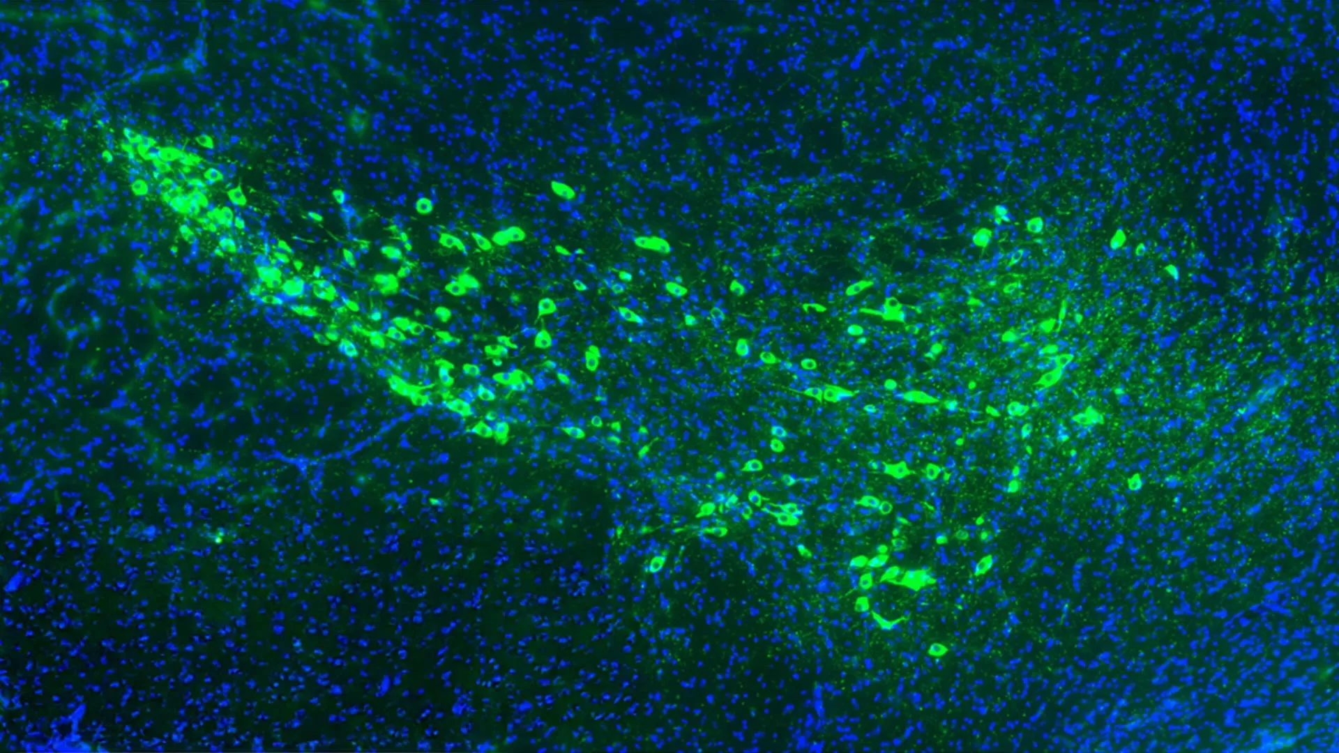
Yale School of Medicine researchers have identified two proteins on neuron surfaces that appear to help Parkinson’s-related damage spread through the brain. The finding matters because blocking those proteins in mice reduced signs of disease progression, pointing to a possible route for treatments aimed at the disease process rather than symptoms alone.
The study, published in Nature Communications, focused on misfolded α-synuclein, a toxic form of a protein that builds up in Parkinson’s disease. Yale said the work points to mGluR4 and NPDC1 as membrane proteins that can carry misfolded α-synuclein into otherwise healthy brain cells.
Parkinson’s disease is a progressive neurological disorder in which brain cells are damaged and die over time. According to the Parkinson’s Foundation, about 1.1 million people in the United States live with the disease, and nearly 90,000 new cases are diagnosed each year.
How the proteins were identified
Yale said researchers tested whether misfolded α-synuclein attaches to proteins on the outside of cells, a possible step in its movement from one neuron to another. The team created 4,400 groups of cells, with each group engineered to display a different surface protein.
Most of those proteins did not interact with misfolded α-synuclein, according to the researchers. Sixteen did bind to it, including mGluR4 and NPDC1, which are found on dopamine-producing neurons in the substantia nigra, a brain region heavily affected in Parkinson’s disease.
The researchers reported that mGluR4 and NPDC1 did more than bind the toxic protein. Yale said the two proteins helped move misfolded α-synuclein into cells, making them candidates for explaining how Parkinson’s pathology can pass between neurons.
Mouse experiments showed reduced disease signs
The team then tested the proteins’ role in animals. Yale said researchers genetically engineered mice so either mGluR4 or NPDC1 no longer worked, then exposed the animals to misfolded α-synuclein.
Normal mice developed brain accumulations of the toxic protein and showed Parkinson’s-like symptoms, according to Yale. Mice lacking functional mGluR4 or NPDC1 did not show the same disease pattern after exposure.
In a separate mouse model of Parkinson’s disease, removing the gene for either protein reduced symptom progression and lowered the risk of death, Yale said. The researchers concluded that mGluR4 and NPDC1 appear to work together in transporting misfolded α-synuclein into neurons, at least in mice.
A possible target, not a treatment yet
Stephen Strittmatter, senior author of the study and chair of neuroscience at Yale School of Medicine, said understanding how α-synuclein enters neurons could help researchers find ways to slow disease spread. Yale quoted him describing misfolded α-synuclein as the “pathologic hallmark of Parkinson’s disease.”
Current Parkinson’s treatments mainly address symptoms, Yale said, including movement problems such as tremors, impaired balance and slowed motion. The new findings suggest that blocking the entry route for misfolded α-synuclein could be explored as a disease-slowing strategy.
The results remain preclinical. Yale presented the work as a potential target for future therapies, not evidence that a drug is ready for patients.
This story draws on original reporting from ScienceDaily.