
Retina study finds linked pathways that may aid low-light vision
Yale researchers report that electrical connections link retinal pathways once thought to work separately, with one cell type helping coordinate weak visual signals.
By Priya Raghavan · Science Reporter
3 min read
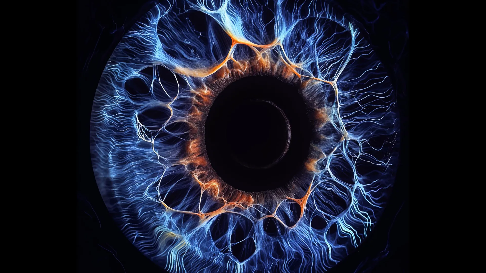
Researchers at Yale School of Medicine have identified an electrical communication system in the retina that links visual pathways previously thought to operate largely on their own. The finding, published in Neuron, may help explain how the eye detects faint contrast and small objects, especially when visual signals are weak.
The study focuses on bipolar cells, neurons that receive information from the retina’s light-sensing rods and cones. According to Yale School of Medicine, those cells sort visual input into more than a dozen channels that handle features such as color, contrast, shape and vision in bright or dim light.
Scientists have treated those channels as mostly separate routes for information moving from the eye toward the brain, the Yale team said. The new work found that many of the channels are tied together by electrical synapses, also known as gap junctions, in both mouse and human retinas.
Yao Xue, a postdoctoral fellow in Yale’s Department of Ophthalmology and Visual Science and first author of the study, said the channels can still carry distinct visual features while also sharing signals through underlying electrical circuitry.
A coordinating cell type
Neurons can communicate through chemical synapses, which use neurotransmitters, or through electrical synapses, which pass currents directly between cells. Yale said bipolar cells had generally been understood as relying mainly on chemical signaling.
In the experiments, the researchers stimulated individual bipolar cells and measured activity in nearby cells. The study found that activating one bipolar cell could trigger neurotransmitter release across multiple bipolar cell types, showing that activity was not confined to a single visual pathway.
The team also identified one bipolar cell type, called BC6, as a central coordinator in the circuit. Z. Jimmy Zhou, the Marvin L. Sears Professor of Ophthalmology and Visual Science at Yale and the study’s principal investigator, said the findings challenge the assumption that bipolar cell types are mostly autonomous.
According to the researchers, signals beginning with BC6 spread through the retinal circuit in an organized hierarchy. Xue said BC6 acts as a “commander” that helps relay signals toward downstream targets.
Seunghoon Lee, a research scientist at Yale and a co-corresponding author, said the integration could be useful when a visual signal is weak and would otherwise be divided among separate channels. Yale said that kind of cooperation may improve detection of low-contrast signals or very small objects.
Intact mouse and human retinas
To map the circuit, the Yale team combined imaging with electrical recordings. The researchers used advanced imaging to observe neurotransmitter activity while stimulating particular cells and recording responses from neighboring cells.
Yale said studying bipolar cells has been difficult because they sit deep in the retina. Earlier work often involved slicing retinal tissue to reach the cells, a step that can disturb the circuits being studied.
For this study, the researchers used a dual patch clamp method in intact mouse retinas, stimulating selected bipolar cell types with electrodes while recording from nearby cells. Zhou said the systematic recordings reflected Xue’s doctoral work and required unusual electrophysiological skill.
The team then performed related experiments on intact human retinas obtained through Yale’s Department of Pathology Legacy Tissue Donation Program. According to Yale, the work represents the first experiments of this type conducted in an intact human retina.
The researchers said the findings could affect more than vision research because the retina is part of the central nervous system. Yale said a clearer view of retinal circuitry may also help scientists study diseases that damage the eye, including macular degeneration, glaucoma and congenital night blindness.
Lee said the project began without a narrow hypothesis and revealed a basic mechanism for visual processing. The study is titled “A hierarchical electrical synaptic circuit mechanism for integrative parallel visual processing in the retina.”
This story draws on original reporting from ScienceDaily.