
Gut toxin’s route into colon cells identified in cancer-linked study
Johns Hopkins-led researchers found that a Bacteroides fragilis toxin binds claudin-4 before damaging colon cells, and blocked it in mice with a decoy protein.
By Priya Raghavan · Science Reporter
3 min read
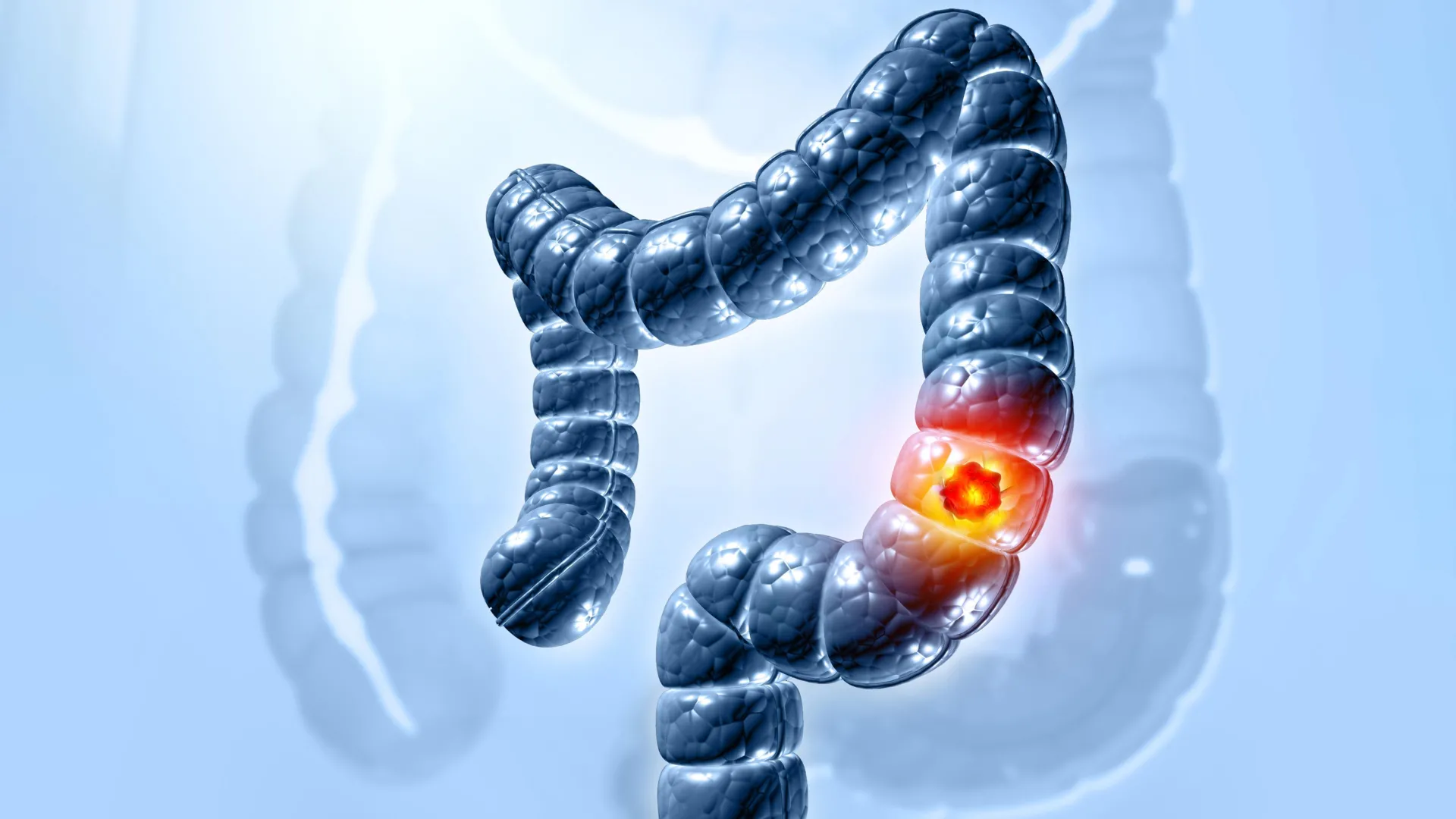
A Johns Hopkins-led team has identified how a toxin from a common gut bacterium gets into position to damage colon cells, a finding tied to research on colorectal cancer. Johns Hopkins Medicine said the work also produced a decoy protein that blocked the toxin’s effects in mice.
The study, published in Nature, focused on BFT, a toxin made by certain strains of Bacteroides fragilis. According to Johns Hopkins Medicine, those strains can inflame the colon and promote tumor growth, although the bacterium is found in up to 20% of healthy people.
Researchers at the Johns Hopkins Kimmel Cancer Center Bloomberg-Kimmel Institute for Cancer Immunotherapy and the Johns Hopkins University School of Medicine led the multi-institutional work. The National Institutes of Health supported the research in part, according to Johns Hopkins Medicine.
How the toxin reaches its target
Johns Hopkins Medicine said earlier work from the laboratory of senior author Cynthia Sears showed that BFT cuts E-cadherin, a protein involved in maintaining the colon’s protective barrier. That prior Nature Medicine study also linked the toxin’s activity to colon tumor formation.
A key problem remained: BFT did not appear to attach directly to E-cadherin. The new study found that the toxin first binds to claudin-4, a host protein, before it can harm colon cells, Johns Hopkins Medicine said.
Sears, a Bloomberg-Kimmel professor of cancer immunotherapy and professor of medicine at Johns Hopkins, said in the university’s announcement that researchers had tried several times to find the receptor. She said understanding bacterial toxins may lead to new detection and treatment approaches for related diseases, including diarrhea, colorectal cancer and bloodstream infections.
CRISPR screen points to claudin-4
Maxwell White, an M.D./Ph.D. candidate in the Sears lab, led a genomewide CRISPR screen with the laboratory of Matthew Waldor at Harvard Medical School, according to Johns Hopkins Medicine. The researchers disabled genes in colon epithelial cells one at a time to see which were needed for BFT to act.
Claudin-4 emerged as the strongest signal in that screen, Johns Hopkins Medicine said. When researchers removed claudin-4, BFT no longer attached to the cells and E-cadherin was not cut.
White said in the announcement that it took time to make the assay work and confirm the method, but claudin-4 stood out once the screen was completed. Johns Hopkins Medicine said the result was unexpected because many researchers had anticipated a signaling protein rather than a claudin protein.
To confirm the interaction, the Johns Hopkins group worked with structural biologists F. Xavier Gomis-Rüth and Ulrich Eckhard at the Molecular Biology Institute of Barcelona. Johns Hopkins Medicine said laboratory biophysical experiments showed BFT and claudin-4 form a tightly bound one-to-one complex.
Decoy protein tested in mice
The team then tested the finding in living systems through work with Min Dong’s laboratory at Harvard Medical School, including Kang Wang and colleagues, Johns Hopkins Medicine said. Researchers made a soluble form of claudin-4 that acted as a decoy, displaying the parts of the receptor recognized by BFT.
According to Johns Hopkins Medicine, the toxin bound to the decoy instead of colon cells, protecting mice from colon damage caused by BFT. White said the approach could be refined using small molecules or other biologic treatments with better drug-like properties.
Johns Hopkins Medicine said the researchers still have not captured the exact experimental structure showing how BFT and claudin-4 fit together. The university said current artificial intelligence modeling tools, including AlphaFold, did not fully resolve that interaction.
The paper listed additional authors from Johns Hopkins and Harvard Medical School. Johns Hopkins Medicine said the work also received support from the Bloomberg-Kimmel Institute for Cancer Immunotherapy, Janssen Research and Development, Cancer Research UK, the Howard Hughes Medical Institute and NIH grants.
This story draws on original reporting from ScienceDaily.