
Researchers build 3D archive of rare vaquita skeleton
Florida Atlantic University researchers used CT imaging to preserve a detailed digital record of the critically endangered porpoise.
By Tom Brennan · Health & Medicine Correspondent
3 min read
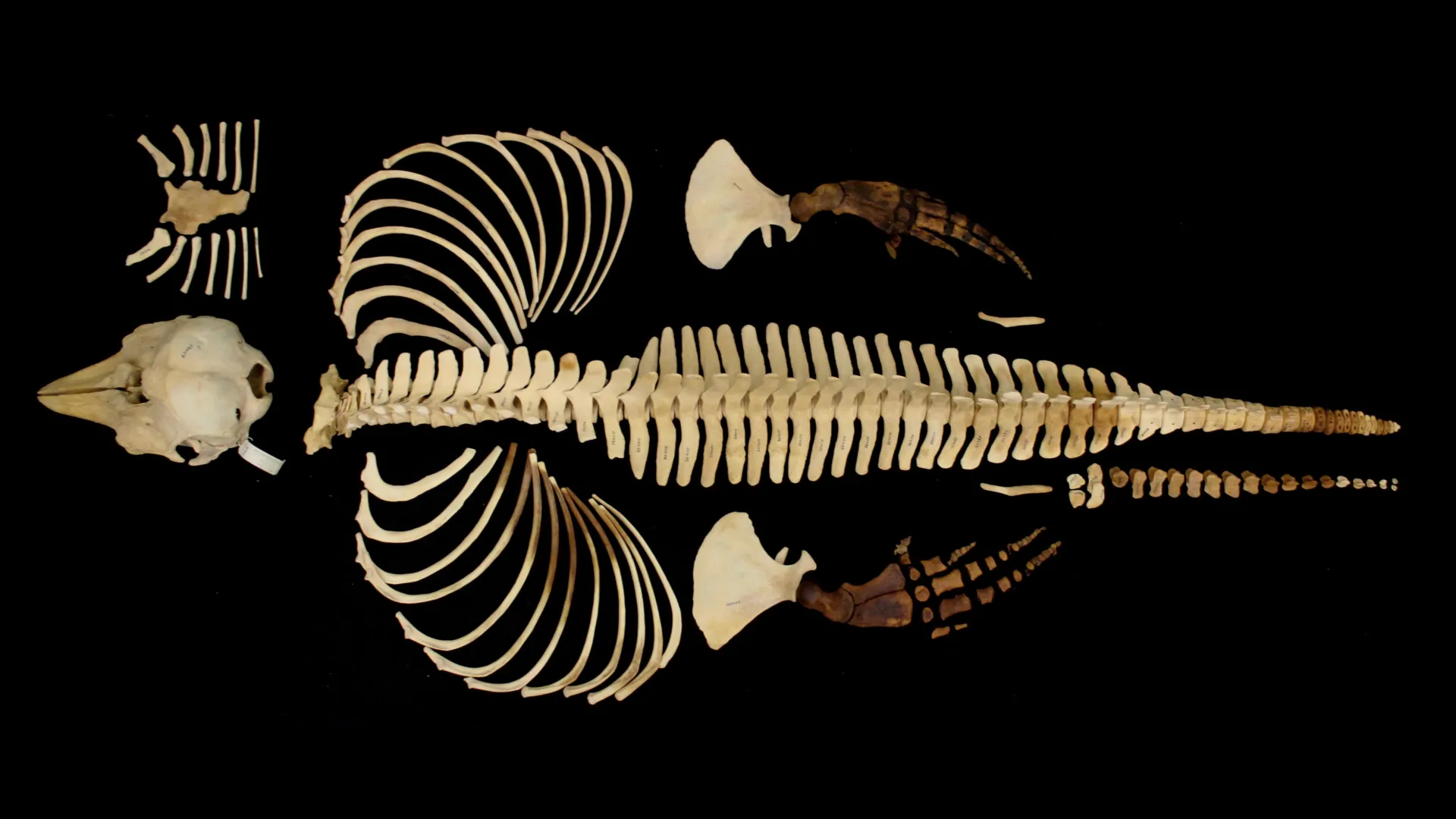
Researchers have created detailed 3D models of a vaquita skeleton, preserving anatomical data from one of the world’s most endangered marine mammals. Florida Atlantic University said the open digital archive could support research, education and conservation awareness as the species nears extinction.
The work, published in Marine Mammal Science, centered on a complete female vaquita skeleton held by the San Diego Natural History Museum. According to Florida Atlantic University, the specimen was collected in the 1960s and donated to the museum in 1966.
The vaquita, or Phocoena sinus, lives only in shallow waters in the northern Gulf of California, Florida Atlantic University said. The porpoise is about 5 feet long and is the smallest cetacean, the group that includes whales, dolphins and porpoises.
Florida Atlantic University said only an estimated handful of vaquitas remain in the wild. The university said the decline has been driven mainly by accidental capture in gillnets, especially nets used illegally to catch totoaba, a fish sought for its swim bladder on black markets.
Scanning a rare specimen
The research team included scientists and specialists from Florida Atlantic University, the San Diego Natural History Museum, SeaWorld California and NOAA Fisheries. Jamie L. Knaub of FAU was the paper’s first author, and Marianne E. Porter, a professor in FAU’s Department of Biological Sciences, was senior author.
According to the university, the team used medical CT scans, micro-CT scans and digital photography to record the skeleton at different scales. The process produced thousands of cross-sectional images, which researchers used to separate individual bones digitally and rebuild them as interactive 3D models.
Micro-CT imaging allowed the team to capture anatomical structures measured in microns, smaller than the width of a human hair, FAU said. The resulting models can be rotated, enlarged and examined from multiple angles without handling the original skeleton.
Knaub said in the university’s announcement that combining advanced imaging with open data sharing protects a rare record of the vaquita and makes it available beyond the institutions that hold the specimen. Knaub said the models also can be used to make accurate replicas for museums, classrooms and education programs.
Open access for researchers and classrooms
FAU said the skeleton’s rarity and fragility limit direct study and public display. To widen access, the team placed the 3D models on MorphoSource, an online repository that allows free access to digital specimen data.
Porter said the project required imaging at several levels, from whole-bone structure to microscopic internal detail. She said the combined methods produced a layered dataset that keeps the specimen’s anatomy intact in digital form.
Tricia L. Meredith, a co-author and director of research for FAU’s on-site lab schools, said the Berlin Family Bioimaging Lab’s micro-CT systems and data-processing expertise were central to turning raw scans into usable models.
The research was supported by FAU’s School of Environmental, Coastal, and Ocean Sustainability, the Joshua M. Berlin Research Gift, FAU Laboratory Schools and SeaWorld California, according to the university.
This story draws on original reporting from ScienceDaily.