
Mini brain models show varied Alzheimer’s drug responses
Johns Hopkins researchers say patient-derived organoids may help identify Alzheimer’s subgroups more likely to respond to specific treatments.
By Priya Raghavan · Science Reporter
3 min read
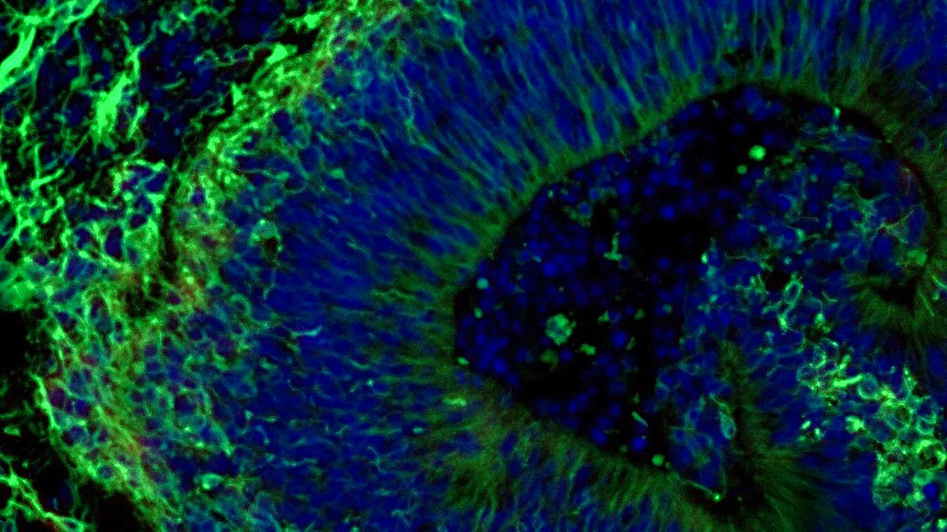
Lab-grown brain tissue made from Alzheimer’s patients’ cells showed different molecular reactions to a commonly used antidepressant, Johns Hopkins Medicine reported. The findings point to a possible path toward more tailored treatment for psychiatric symptoms tied to Alzheimer’s disease.
The study, published in Alzheimer’s & Dementia: The Journal of the Alzheimer’s Association, examined brain organoids, small three-dimensional tissue models grown in the lab. Johns Hopkins said the work also found that tiny particles released by the organoids may contain clues useful for diagnosing Alzheimer’s disease or assessing its progression.
Alzheimer’s disease has no cure and is the most common form of dementia, affecting more than 7 million Americans, according to Johns Hopkins Medicine. Selective serotonin reuptake inhibitors, or SSRIs, are often used to treat anxiety, depression and agitation in people with the disease, but Johns Hopkins researcher Vasiliki Machairaki said patients respond unevenly to those drugs.
Patient cells turned into hindbrain tissue
The research team used blood samples from people with Alzheimer’s disease who were enrolled through the NIH-funded Johns Hopkins Alzheimer’s Disease Research Center, Johns Hopkins said. Scientists reprogrammed blood cells into induced pluripotent stem cells, which can develop into many cell types.
Using cells from Alzheimer’s patients and healthy participants, the team grew organoids resembling the hindbrain, a region involved in functions such as breathing, sleep and heart rate. The organoids included neurons that produce serotonin, the chemical pathway targeted by SSRIs.
Johns Hopkins said the study included hundreds of organoids representing individual Alzheimer’s patients and healthy controls. Machairaki said it may be among the largest brain organoid studies yet conducted in Alzheimer’s research.
Antidepressant response varied by sample
The researchers reported that organoids grown from Alzheimer’s patients’ cells showed molecular differences from organoids made from healthy participants’ cells. Those differences involved proteins linked to brain-cell communication, inflammation and Alzheimer’s-related pathways, according to Johns Hopkins.
The team then exposed the organoids to escitalopram oxalate, an SSRI widely prescribed as an antidepressant. Some Alzheimer’s-derived organoids showed increases in proteins connected to serotonin signaling and communication between neurons, while others showed little or no response, Johns Hopkins said.
Machairaki said the model could eventually help researchers identify patient subgroups whose biology suggests they are more likely to benefit from certain drugs. She described the work as an early step toward more precise, targeted treatment strategies.
Released particles may carry disease signals
The study also examined extracellular vesicles, small particles released by cells that carry proteins and other cellular information. Johns Hopkins said vesicles from the organoids contained proteins involved in neuron communication, memory and neurotransmitter release.
In vesicles from Alzheimer’s-derived organoids, the researchers found lower levels of RAB3A, NSF and ATCAY, proteins involved in normal signaling between brain cells. After escitalopram treatment, some samples showed increases in proteins tied to serotonin signaling and synaptic pathways, according to the study summary from Johns Hopkins Medicine.
Machairaki said those vesicles could eventually help indicate which patients are more likely to respond to a given treatment. She also said future work will focus on more complex organoids that include immune cells and vessel-like networks, which could make the models closer to human brain tissue.
The study received support from the National Institutes of Health, the Paul G. Allen Frontiers Foundation and the Richman Family Precision Medicine Center of Excellence in Alzheimer’s Disease at Johns Hopkins University. Johns Hopkins said no authors declared a related conflict of interest under university policies.
This story draws on original reporting from ScienceDaily.