
Malaria cell-entry structure reveals possible route to new drugs
Columbia researchers mapped a short-lived malaria invasion complex and designed a mini-protein that blocked parasite entry into red blood cells.
By Lucas Ferreira · Science & Environment Writer
3 min read
Columbia University researchers have captured a high-resolution structure of the malaria parasite’s “moving junction,” a short-lived complex the parasite uses to enter human red blood cells, and used the structure to design a mini-protein that blocked invasion in lab tests. The findings matter because malaria kills roughly 600,000 people a year, mostly young children in sub-Saharan Africa, while the parasite is becoming resistant to leading drugs, according to Columbia University Irving Medical Center.
The work, published in Cell, addresses a basic question in malaria biology that has remained unresolved for decades: what the moving junction does during the first step of infection. Columbia said the complex forms, acts and disappears in less than 60 seconds, making it hard to study in detail.
A fast event caught midstream
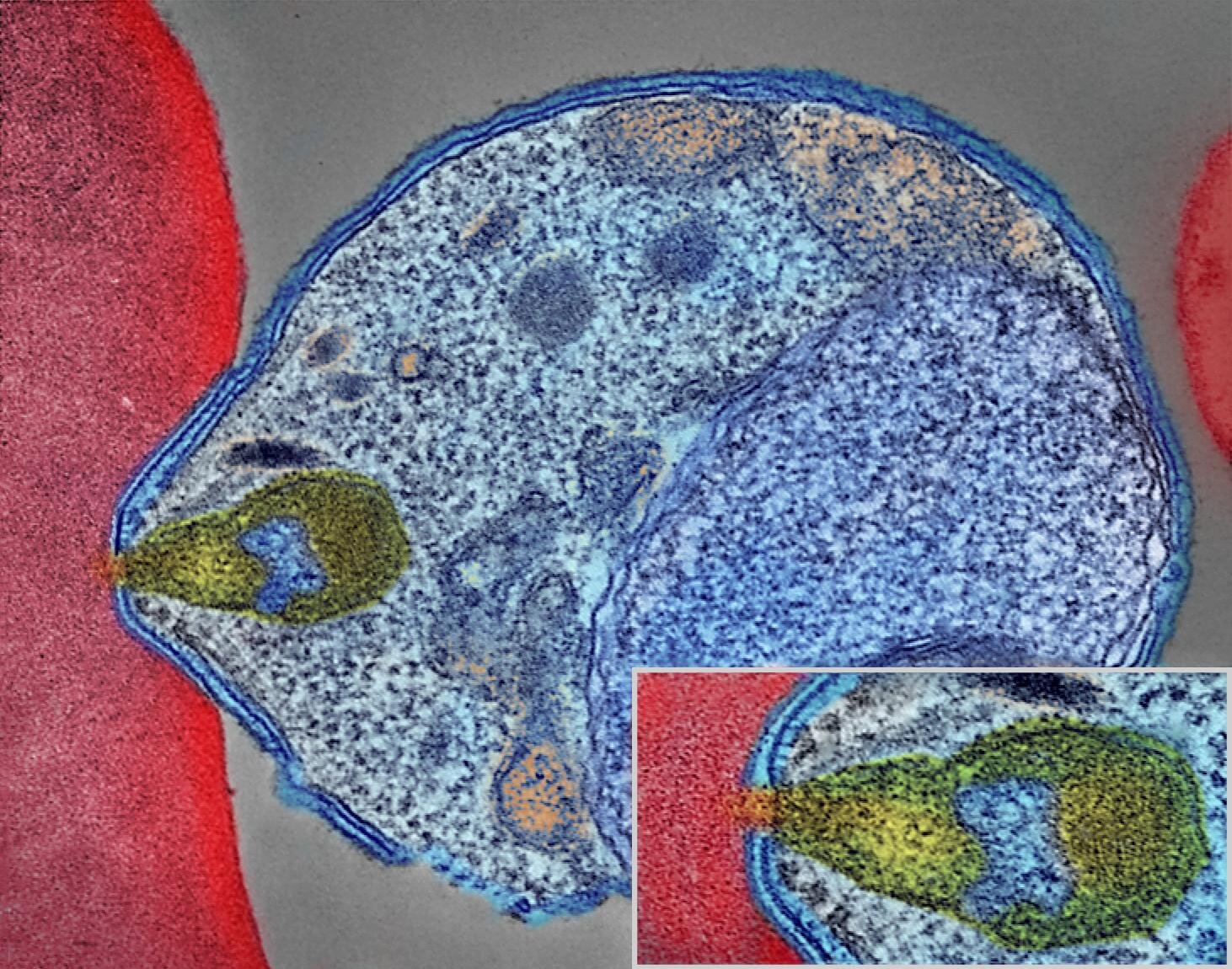
Scientists first saw the moving junction in 1978 as a thickened region of membrane where a parasite meets a host cell, according to Columbia. Researchers later identified four parasite proteins that make up its core building block — AMA1, RON2, RON4 and RON5 — and showed that they are required for invasion.
The Columbia team stalled parasites as they were entering red blood cells by using a compound that stops the parasite’s internal motor while allowing the junction to form, the university said. The researchers then removed the intact AMA1-RON complex from the cell and studied it with cryo-electron microscopy, which freezes molecules and images them with an electron beam.
That approach produced a three-dimensional view of the complex. Columbia reported that the structure resembled a sailboat, with AMA1 above the cell surface and the RON proteins pressed against the host membrane.
A machine that reshapes membrane
The structure changed the researchers’ understanding of how the parasite enters a cell, according to the Cell paper. Columbia said the moving junction appears to act as a membrane-remodeling machine rather than a passive ring through which the parasite pulls itself.
The team found positively charged anchors on the surface facing the host membrane, along with short helical segments that insert into the membrane, Columbia said. Those features are associated with cellular machinery that bends or reshapes membranes.
To test that idea, the researchers made versions of the parasite’s wedge-like helices and added them to artificial membrane bubbles, according to Columbia. The membranes thinned and developed holes, while weakened versions of the helices did not damage the bubbles.
Chi-Min Ho, assistant professor in Columbia’s Department of Microbiology and Immunology and senior author of the study, said the team had known the structure was essential for invasion but lacked a clear mechanism. Meseret Haile, the study’s first author and a Ph.D. candidate in Ho’s lab, said the results show a machine built to reshape the host cell membrane.
Mini-protein blocks entry
The structure also showed how AMA1 binds its partner protein, a contact Columbia said helps hold the junction together. Using that map and a machine-learning protein-design tool, the researchers designed a mini-protein meant to disrupt the interaction.
Columbia said the best candidate blocked parasites from invading red blood cells in a dose-dependent manner and did not affect cells that were already infected. The university said that result indicates the mini-protein acted by preventing entry rather than by causing broad toxicity.
The candidate remains a proof of concept and would need further refinement before any testing in people, according to Columbia. The researchers said the same structural information may also help explain how leading anti-malaria antibodies work and could inform vaccine design.
The study, “Structural basis for host membrane binding and remodeling by invading malaria parasites,” was published in Cell.
This story draws on original reporting from Phys.org.