
Fat-loaded immune cells linked to faster MS progression
A study of donated brain tissue ties lipid-filled microglia to severe multiple sclerosis and possible future biomarkers.
By Lucas Ferreira · Science & Environment Writer
3 min read
Researchers in the Netherlands have identified fat-filled immune cells in the brain that are associated with more severe multiple sclerosis, according to the Netherlands Institute for Neuroscience. The findings matter because they could help explain why MS advances quickly in some patients and more slowly in others.
The work, led by Daan van der Vliet with researchers from the Netherlands Institute for Neuroscience, Leiden University and Utrecht University, was published in Nature Neuroscience. The team studied donated brain tissue from 28 deceased people with MS through the Netherlands Brain Bank, according to the institute.
MS attacks myelin, the fatty layer that protects nerve fibers in the brain and spinal cord, the institute said. As that covering breaks down, patients can develop neurological symptoms that include trouble walking and impaired vision.
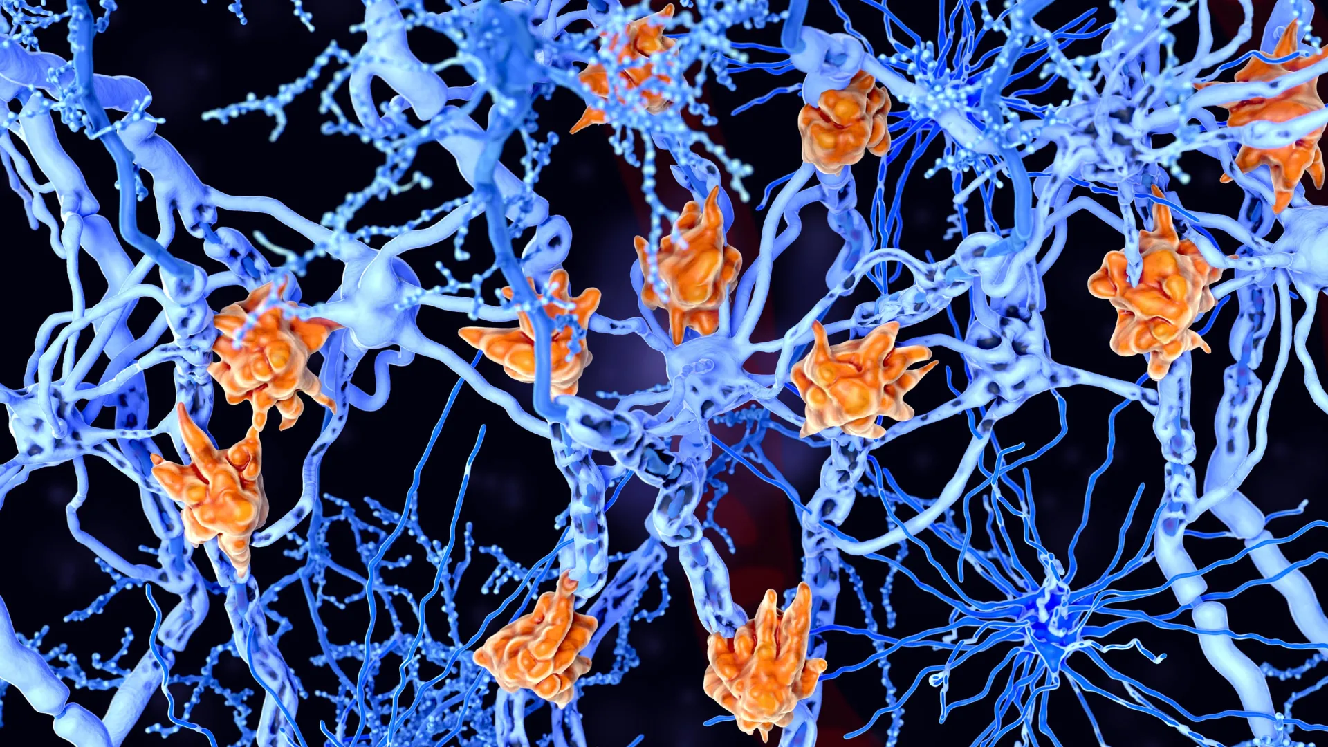
Cleanup cells under strain
The study focused on microglia, immune cells in the brain that help clear damaged material and support repair, according to the research team. In tissue from patients with rapidly progressing MS, the scientists found many microglia packed with lipid droplets after taking in damaged myelin.
Researchers call these cells “foamy microglia” because of their appearance. Van der Vliet said patients with high numbers of these cells more often had a severe course of disease, according to the Netherlands Institute for Neuroscience.
The team’s interpretation is that the cells may begin as part of the brain’s repair response. Van der Vliet said the microglia appear to be trying to clear injury-related debris, but can become overloaded and lose their ability to aid recovery.
The study also found molecular differences between MS lesions that contained foamy microglia and lesions that did not, according to the institute. Lesions with these cells showed enrichment of certain fats tied to persistent inflammatory activity.
A wider view of MS damage
Inflammation has long been treated as a central driver of MS progression, the researchers said. Van der Vliet said the new findings point to a process in which failed cleanup and repair may intensify inflammation and interfere with recovery.
To study the lesions, the scientists combined analyses of gene activity, proteins and lipids in affected brain tissue, according to the Netherlands Institute for Neuroscience. Van der Vliet said the work depended on pairing advanced mapping methods with the Netherlands Brain Bank’s detailed classification of brain pathology.
The published paper is titled “Foamy microglia link oxylipins to disease progression in multiple sclerosis.” Its authors include Van der Vliet and colleagues from the participating Dutch institutions, with the journal listing the work under DOI 10.1038/s41593-026-02302-3.
Possible biomarkers and treatment targets
The researchers said the findings could help in the search for biomarkers that flag patients at higher risk of rapid decline. They reported evidence that some fats associated with foamy microglia may also be detectable in cerebrospinal fluid, though the institute said future studies would need to confirm that possibility.
If confirmed, those molecules could help doctors identify patients whose disease is likely to worsen faster and guide treatment choices earlier, according to the research team. The institute said the findings also align with experimental efforts to target fat metabolism and chronic MS lesion growth.
Several related treatment approaches are being tested in clinical studies conducted in collaboration with Roche, according to the Netherlands Institute for Neuroscience. The research was supported by the Institute for Chemical Immunology and the Institute for Chemical NeuroScience, both Gravitation programs, the institute said.
This story draws on original reporting from ScienceDaily.