
Study maps DNA-folding proteins that shape antibody diversity
Michigan State researchers report that two cohesin variants help immune cells assemble antibody genes by controlling long-range DNA loops.
By Tom Brennan · Health & Medicine Correspondent
3 min read
Michigan State University researchers have identified how two related DNA-organizing proteins help immune cells assemble antibody genes. The finding matters because antibody diversity depends on immune cells joining genetic segments that can sit far apart on the same chromosome.
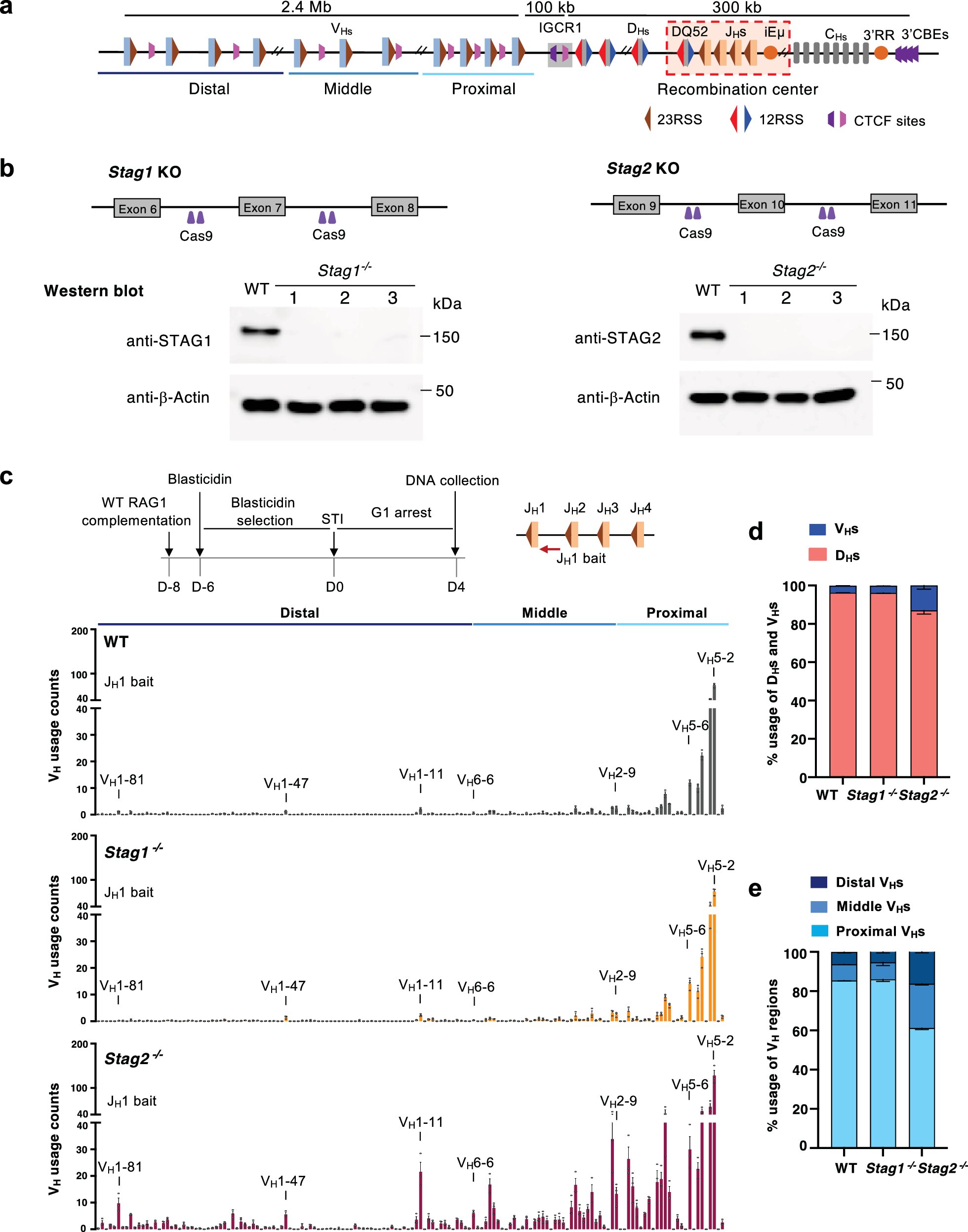
The work, published in Nature Communications, focuses on cohesin complexes containing the proteins STAG1 and STAG2. According to the MSU team, the two proteins do different jobs during the process that builds antibody genes, despite being closely related parts of the same chromosome-folding machinery.
Antibodies are produced from a shared genome, but the immune system needs many versions to respond to bacteria, viruses and other pathogens. Immune cells create that variety by rearranging gene segments known as V, D and J segments, the researchers said.
Those segments are not lined up side by side along DNA. Cells must bring distant pieces into contact with enough control to make useful antibody genes, a process tied to the three-dimensional folding of chromosomes.
Yu Zhang, an assistant professor in MSU’s Department of Microbiology, Genetics, and Immunology and senior author of the study, said the central problem has been how cells accurately connect DNA segments separated by long stretches of genetic code. Zhang said the study shows that different versions of chromosome-organizing machinery can control that process in different ways.
STAG1 and STAG2 split the work
The researchers studied cohesin, a protein complex that helps fold DNA into loops through a process known as loop extrusion. In the antibody gene system examined by the team, STAG2 restrained long-range looping at an early stage, while STAG1 supported the distant DNA contacts needed for assembly.
According to the study, when STAG2 was absent, STAG1 produced longer DNA loops and allowed antibody gene segments to interact earlier than they otherwise would. The team also found that STAG1 works with CTCF, another chromosome-organizing protein, to form long-distance interactions efficiently.
At later stages of antibody gene assembly, the MSU researchers said both STAG1-containing and STAG2-containing cohesin complexes contribute to bringing many V segments across long DNA distances into the process. That coordination helps generate a broader range of antibodies.
Jianrong Wang, an MSU associate professor who led computational data integration and bioinformatics analysis for the study, said combining computational and laboratory approaches is becoming central to understanding how genomes reorganize in three-dimensional space and how those changes affect cell function.
Links to disease research
The study centers on antibody formation, but MSU said the same chromosome-organizing proteins help maintain genome organization more broadly. Defects in these proteins have been associated with developmental disorders and several cancers, including diseases that affect blood and immune cells.
The researchers said the findings could help clarify how abnormal immune-cell development contributes to leukemia and lymphoma. They did not report a new treatment, but described a mechanism that may help explain how mistakes in DNA organization lead to disease.
The paper, titled “Context-dependent regulation of IgH V(D)J recombination by cohesin-STAG1 and cohesin-STAG2,” lists Fujung Chang and colleagues as authors. Its DOI is 10.1038/s41467-026-74012-0.
This story draws on original reporting from Phys.org.