
Cryo-EM reveals how gum disease bacterium twists through tissue
Yale researchers mapped Treponema denticola’s flagella at near-atomic detail, showing a two-part structure tied to its movement.
By Priya Raghavan · Science Reporter
3 min read
Yale researchers have used cryo-electron microscopy to show how a major gum disease bacterium builds the machinery that helps it move through tissue. The work matters because Treponema denticola is one of the Red Complex bacteria linked to chronic periodontal disease, which the Centers for Disease Control and Prevention estimates affects about 40% of U.S. adults over 30.
The findings were reported by Yale University and published in Nature Communications. The study focuses on the flagella of Treponema denticola, the hairlike structures that power the bacterium’s corkscrew motion.
According to Yale, Treponema denticola belongs to the spirochetes, a group of long, thin bacteria known for spiral shapes and the ability to move through thick biological material. Jiaqi Wang, a postdoctoral associate in Jun Liu’s lab at Yale’s Microbial Sciences Institute, said the field had known that flagella drive that motion, but had not explained how the structures assemble and function in this bacterium.
A closer look at bacterial movement
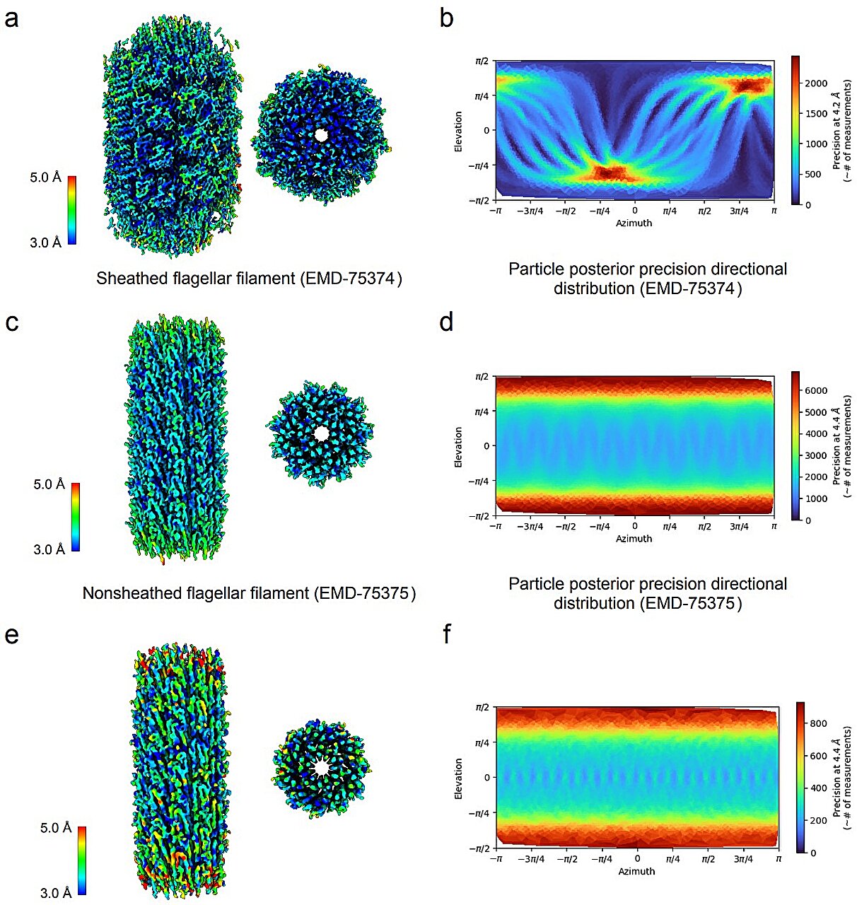
The Liu lab used cryo-EM, a form of electron microscopy that can image biological structures at near-atomic resolution, to examine the bacterium’s flagellar filament. Yale said the analysis showed an asymmetric design that differs from flagellar structures seen in other bacteria.
The Nature Communications paper describes a filament architecture built from two major parts: a central core and an outer sheath. Yale said that arrangement allows the entire spirochete to twist, helping explain how the organism can bore through human cell tissue during infection.
Wangbiao Guo, an associate research scientist and co-first author of the study, said in Yale’s account that the team found a central core and outer sheath rather than a more typical bacterial filament structure. Yale said the discovery came after the researchers constructed a new protein sequence for the bacterium’s flagellar filament.
The study was led from Liu’s lab at Yale’s West Campus. Yale said Wang brought structural biology expertise to the project, Guo moved from engineering into microscopy, and Chuck Sindelar, a cryo-EM specialist at the Yale Center for Research Computing, contributed insight into the asymmetric structure of spirochete flagella.
Potential implications for treatment research
Yale said the work addresses a problem that has challenged researchers for decades: how Treponema flagella are organized and how they generate the bacterium’s movement. Liu, a professor of microbial pathogenesis at the Yale Microbial Sciences Institute, said the imaging advance allowed the team to see how the flagella function rather than only their general shape.
Yale also pointed to its West Campus imaging resources, including 3D fluorescence and optical microscopes, as part of a broader research program aimed at studying cellular structures and dynamics. The university said similar tools have supported recent structural work on Legionella bacteria and COVID-19 defenses.
The paper, “Asymmetric architecture and adaptation of Treponema flagella,” was published in Nature Communications in 2026. Yale said the combination of microscopy and cross-disciplinary research could help guide future work on possible orthodontic treatment methods.
This story draws on original reporting from Phys.org.