
Wasteosomes in neurodegenerative diseases may signal poor brain clearance
A University of Barcelona-led study found more wasteosomes in diseased brains, pointing to a possible shared clearance failure.
By Tom Brennan · Health & Medicine Correspondent
3 min read
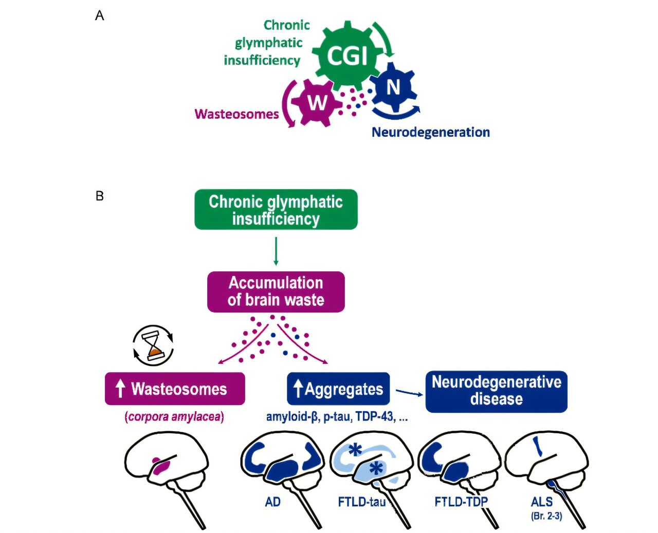
A University of Barcelona-led study of wasteosomes in neurodegenerative diseases found that these small waste-filled structures were more common in brain samples from people with Alzheimer’s disease, amyotrophic lateral sclerosis and frontotemporal lobar degeneration. The findings point to a possible shared problem in the brain’s waste-removal system, which researchers say could help explain how several neurodegenerative conditions develop or worsen.
The study was published in Acta Neuropathologica Communications. According to the University of Barcelona, the work was led by Jordi Vilaplana of the university’s Faculty of Pharmacy and Food Science, its Institute of Neurosciences and CIBERNED, together with Laura Molina-Porcel of the Neurological Tissue Bank Biobank at Hospital Clínic de Barcelona–IDIBAPS.
What are wasteosomes in neurodegenerative diseases?
Wasteosomes, also known as corpora amylacea, are small structures described by the researchers as containers for waste material in the brain. The University of Barcelona said their buildup may serve as an indirect sign that the glymphatic system, which helps clear waste from the brain, has been underperforming over a long period.
The researchers examined brain tissue from 185 donors. The group included donors with Alzheimer’s disease, ALS and frontotemporal lobar degeneration, as well as donors without a neurodegenerative disease, according to the university.
The analysis found that wasteosomes accumulated repeatedly in certain brain regions in people with disease. The University of Barcelona said those regions were linked to drainage pathways used by the glymphatic system, and the number of wasteosomes was significantly higher in brains from affected donors.
The researchers also reported that the pattern did not match the distribution of the disease-specific abnormal proteins seen in each condition. They said that finding supports the idea that wasteosomes may reflect a common mechanism across different neurodegenerative diseases, rather than being a local reaction to protein deposits.
Why the glymphatic system is under scrutiny
The glymphatic system is a brain clearance pathway involved in removing altered proteins and other waste products. According to the research team, if that system works inefficiently, waste can build up in ways that may contribute to the start or progression of neurodegenerative disease.
The University of Barcelona said one possible implication is that wasteosomes could become a biomarker for chronic glymphatic insufficiency. The researchers said current tools for assessing long-running changes in glymphatic function are limited.
If further studies confirm the link, the team said wasteosome-related measures could help identify people at higher risk of neurodegenerative disease or help track disease progression. The findings also support further study of approaches aimed at preserving or restoring glymphatic function, according to the researchers.
The next step, the University of Barcelona said, is to produce direct evidence tying wasteosome accumulation to impaired brain clearance. The researchers said that will require experiments combining measurements of glymphatic function with wasteosome analysis in animal models and, where possible, in human studies.
The team also said future work should examine other neurodegenerative diseases and develop noninvasive imaging tools that can detect wasteosomes or the brain changes associated with them. Marta Riba and Raquel Alsina were listed as first authors, with researchers from the Universitat Politècnica de Catalunya and Qilimanjaro Quantum Tech also taking part.
This story draws on original reporting from Medical Xpress.