
MRI analysis may detect heart attack effects beyond scarred tissue
Researchers say strain analysis on cardiac MRI could reveal subtle ventricular dysfunction after myocardial infarction.
By Tom Brennan · Health & Medicine Correspondent
3 min read
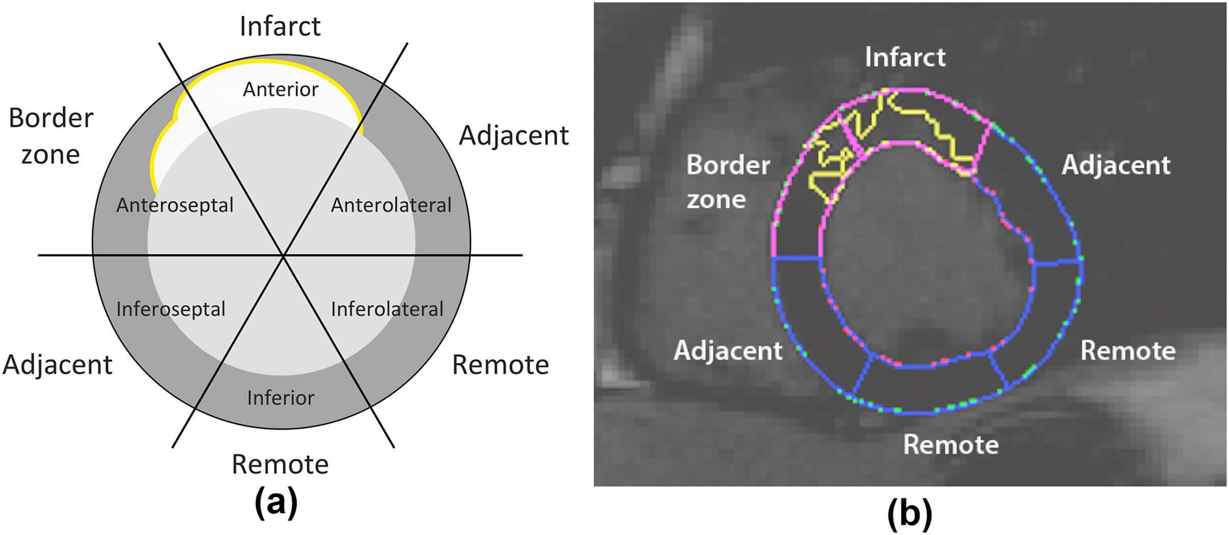
Damage from a myocardial infarction may affect parts of the ventricle that appear distant from the scarred area, according to researchers in Valencia. The finding points to a possible way to spot cardiac decline earlier in patients who survive heart attacks.
The study, reported by the Technical University of Valencia and published in Clinical Radiology, used advanced cardiac magnetic resonance imaging to examine chronic myocardial infarction. Researchers focused on whether regions outside the obvious infarct zone showed functional changes that standard measurements can miss.
The work was conducted by teams from the INCLIVA Health Research Institute, the Clinical University Hospital of València, the University of Valencia, the Universitat Politècnica de València, the Cardiac Imaging Unit of Ascires Biomédico Group and the Ascires-UPV Joint Research Unit.
Why remote dysfunction matters
Myocardial infarction is a major form of ischemic heart disease. The World Health Organization says cardiovascular diseases remain the world’s leading cause of death and account for nearly 18 million deaths each year, with heart attacks making up a substantial share.
Researchers said improved acute treatment has raised survival after heart attacks in Spain and other developed countries. That progress has also left more people living with longer-term complications, including ventricular dysfunction and risk of heart failure.
The Valencia team said identifying dysfunction away from the infarcted tissue could improve prognosis, help detect deterioration before symptoms or more visible structural changes emerge, and support earlier treatment decisions tailored to individual patients.
How the team studied the heart
The researchers used cardiac MRI with feature tracking, a method that measures myocardial strain, or how the heart muscle deforms during the cardiac cycle. According to the team, this can offer a more sensitive view of function than conventional approaches.
Dr. Irene del Canto Serrano, first author of the study, said the aim was to determine whether heart regions not visibly affected by the infarction also show subtle impairment. Del Canto is part of INCLIVA’s Experimental Cardiac Electrophysiology Research Group and is an associate professor in the Department of Electronic Engineering at the University of Valencia.
David Moratal, a professor in the Department of Electronic Engineering at UPV, co-led the research. The team also included Dr. Francisco Javier Chorro Gascó and Dr. Vicente Bodí Peris of INCLIVA, the Hospital Clínico Universitario de València and the University of Valencia, as well as Dr. María Pilar López-Lereu and Dr. José Vicente Monmeneu from Ascires Biomédico Group.
The study built on earlier work in healthy volunteers that established reference values for myocardial strain measured by cardiac MRI, according to Moratal. He said those baseline values allowed the researchers to compare chronic infarction patients against normal ranges and detect changes that would not be apparent using traditional measures such as ejection fraction.
The researchers analyzed cardiac MRI images from patients with chronic myocardial infarction and classified myocardial segments by their relationship to scar tissue, including infarcted, border, adjacent and remote regions. Their report says the method identified functional abnormalities in areas that otherwise appeared healthy.
The paper, “Assessment of remote ventricular dysfunction by cardiac magnetic resonance feature tracking strain in chronic infarction,” carries the DOI 10.1016/j.crad.2025.107205.
This story draws on original reporting from Medical Xpress.