
Mouse study suggests aging ovaries shift toward immune activity
Researchers found post-reproductive mouse ovaries showed inflammation-linked gene activity and immune-cell buildup after follicles were gone.
By Priya Raghavan · Science Reporter
3 min read
Ovaries that have stopped supporting reproduction may remain biologically active in a different way, according to a study in Molecular Human Reproduction. The findings matter because they challenge the view that postmenopausal ovaries are mostly dormant and point to a possible role in inflammation during aging.
The study, by Aubrey Converse and colleagues, examined mouse ovaries across reproductive aging. Researchers reported that after the animals entered a post-reproductive stage, the ovaries showed signs of changing from organs centered on eggs and reproductive hormones into tissue marked by immune activity.
Menopause in humans is defined as 12 straight months without a menstrual period and usually occurs in the 40s or 50s. The mouse equivalent studied here is called oopause, a period after fertility has declined and egg production has ended.
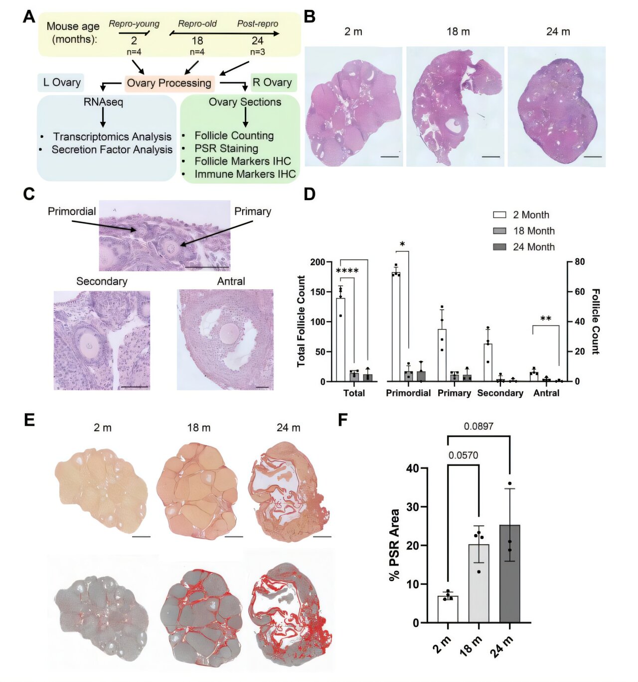
How the study tracked ovarian aging
The researchers used mice at three life stages: reproductively young animals at 2 months old, reproductively aged animals at 18 months old and post-reproductive animals at 24 months old. One ovary from each animal was used to examine structure, while the other was analyzed for gene activity through transcriptomics.
That design let the team compare physical changes in the ovaries with shifts in what cells were doing at the molecular level. The researchers also used existing data sets to identify genes linked to molecules that could signal to other parts of the body.
By the post-reproductive stage, the mice had no remaining follicles, the small sacs that contain immature eggs. The researchers also found that the ovaries had become stiffer, which they linked to a marked increase in collagen.
Immune signals replaced reproductive programs
The largest shift was molecular. According to the study, genes involved in egg production and reproductive hormone synthesis were mostly switched off in the post-reproductive ovaries.
At the same time, genes tied to inflammation, immune responses and white blood cell activation were strongly active. The researchers also observed an influx of immune cells, including T cells, macrophages and large giant cells.
The team reported that the transformed ovaries produced chemical signals that could circulate through the bloodstream. The study suggests those signals may allow the post-reproductive ovary to influence inflammatory processes elsewhere in the body, although the work was conducted in mice.
The authors said the findings argue against treating the ovary as inactive once reproduction ends. Instead, the mouse data suggest the organ may take on an immune-like role after its reproductive function is lost.
Questions for human health
The study does not establish that the same changes occur in human ovaries. Researchers noted that human ovarian samples are difficult to obtain, which is one reason mice were used to build a timeline of ovarian aging.
If future studies find similar patterns in people, the work could shape research into postmenopausal health. The authors suggested that targeting ovarian inflammation might eventually be explored as a way to reduce risks tied to inflammatory and age-related disease in postmenopausal women.
For now, the finding reframes a basic question in reproductive aging: what the ovary does after it stops producing eggs. In mice, the answer from this study is that it keeps changing, with immune signaling becoming a prominent feature.
This story draws on original reporting from Medical Xpress.