
Injectable biomaterial stroke repair study shows gains in mice
Duke researchers report that a hydrogel scaffold recruited immune cells and improved repair markers after stroke in mice.
By Tom Brennan · Health & Medicine Correspondent
3 min read
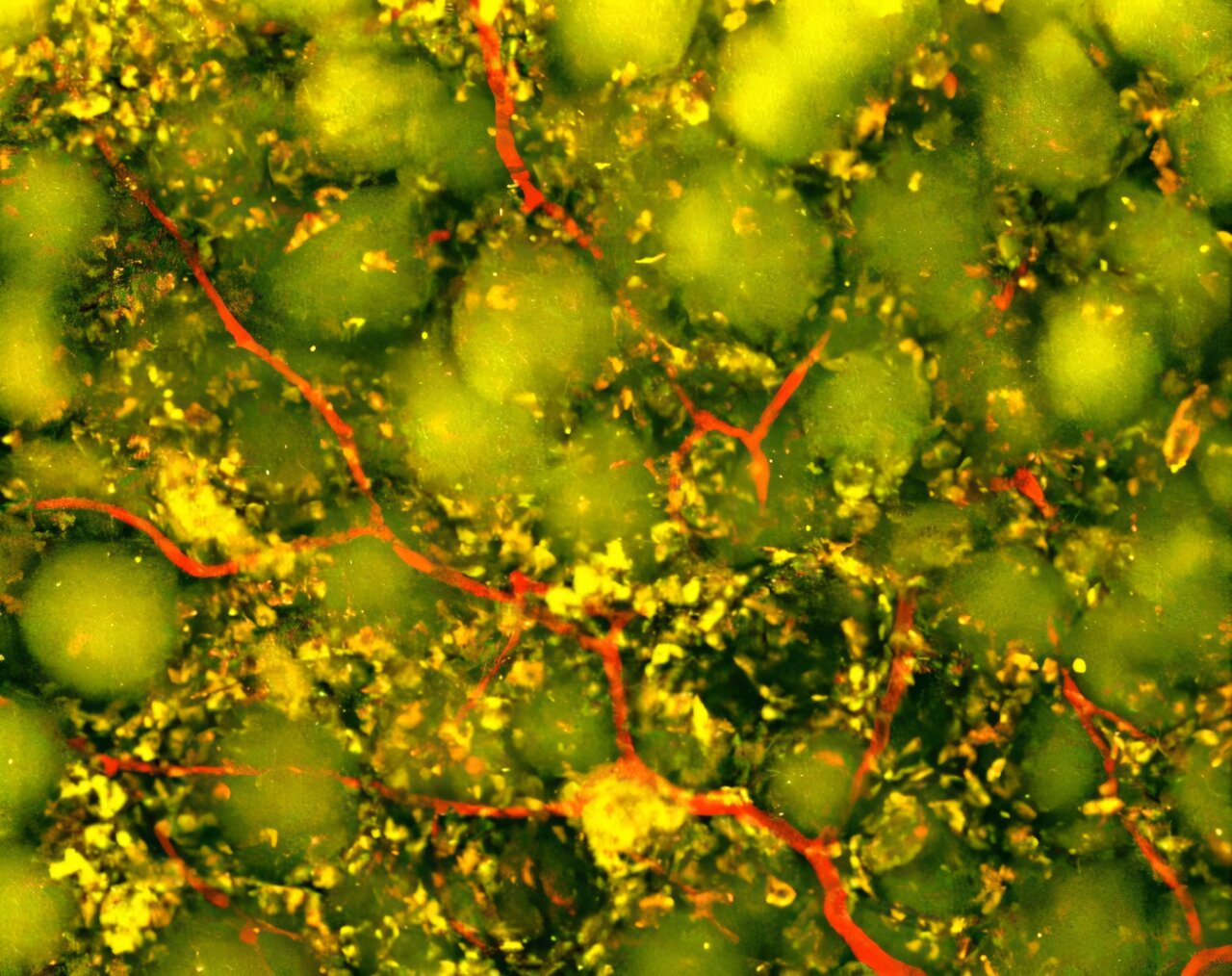
Duke University biomedical engineers say an injectable biomaterial for stroke repair helped turn injury cavities in mouse brains into sites more favorable to healing. The preclinical treatment recruited immune cells, encouraged new blood vessel growth, supported neural remodeling and improved motor performance, according to a study published in Cell Biomaterials.
The work addresses a gap left after ischemic stroke, the type caused by a blood clot. Duke said clot-dissolving drugs and mechanical clot removal can restore blood flow and save threatened brain tissue, but they do not replace tissue that has already died.
Large strokes can leave a cavity where tissue was lost. Duke said rehabilitation can help remaining brain circuits adapt, while the new approach aims to make the damaged space more supportive of biological repair.
How could an injectable biomaterial help after stroke?
The Duke team used microporous annealed particle scaffolds, or MAPS, made from individual hydrogel microparticles. The particles form a porous structure that gives incoming cells a place to enter, interact and build tissue-like networks, according to Duke.
The researchers added extracellular vesicles from astrocytes, star-shaped brain cells involved in normal brain support and injury response. Extracellular vesicles are tiny packages released by cells that can carry proteins, lipids and genetic material to other cells.
Rather than inject the vesicles by themselves, the team chemically attached them to the hydrogel particles. Duke said that design kept the signals in the scaffold so immune cells entering the stroke cavity could encounter them locally.
Immune cells were part of the repair response
The researchers tested vesicles carrying different signaling combinations. Duke said the strongest results came from a combination involving IL-4 and C1q, which drew immune cells into the damaged region, including macrophages and a persistent population of neutrophils.
Neutrophils are often linked to inflammation and tissue injury early after stroke. In this study, Duke said the cells appeared to have a different role later in recovery when they were recruited into the engineered material environment.
When the researchers depleted the neutrophil-rich immune-cell population, blood vessel growth and remodeling of the scaffold were reduced, according to Duke. Shangjing Xin, the study’s lead scientist and a postdoctoral fellow in Tatiana Segura’s laboratory, said the findings suggest neutrophils’ effects after stroke depend on timing, location and surrounding signals.
What changed in the treated mice?
Duke said treated stroke cavities developed blood vessels throughout the scaffold. The researchers also found more axonal fibers in and around the damaged region; axons are the long projections that brain cells use to send signals.
Mice given the optimized scaffold improved on a grid-walking test that tracks forelimb placement errors. By eight weeks, Duke said their performance was statistically indistinguishable from healthy control mice, and the improvement lasted through the study period.
The scaffold itself appeared to be necessary. Duke said extracellular vesicles delivered without the MAP scaffold did not produce comparable vascular repair, suggesting the porous structure and localized signaling were central to the effect.
What happens next?
The results remain limited to mouse models, and the material was injected directly into the damaged brain site. Duke said further work is needed to assess safety, clarify how different immune-cell populations affect recovery and test the method in larger, more clinically relevant stroke models.
The current study used extracellular vesicles from primary rat astrocytes. Duke said Segura’s laboratory is now exploring vesicles made by astrocytes derived from human induced pluripotent stem cells, a possible route toward a more scalable and clinically relevant material source.
This story draws on original reporting from Medical Xpress.