
New imaging method tracks microplastics in living tissue for months
Researchers say photoacoustic imaging can map common plastic particles inside living mice without dissection or chemical labeling.
By Tom Brennan · Health & Medicine Correspondent
3 min read
Researchers have used a noninvasive imaging method to map microplastics deep inside living tissue in fine detail over months, according to a study published in Advanced Science. The work matters because scientists studying possible health effects of microplastics have often had to rely on biopsies or tissue analysis after dissection, limiting what they could observe over time.
The study involved researchers from Kingston University London, University College London and the University of Birmingham. They reported detecting common plastics including polypropylene, used in items such as food containers and coffee cups, and polyethylene, used in single-use plastic bags.
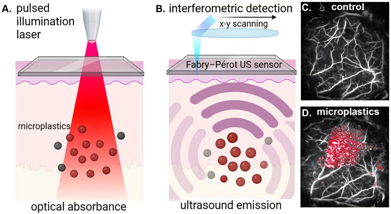
The team used photoacoustic imaging, a technique that sends laser pulses into tissue. According to the researchers, microplastics absorb the light in a distinctive way, producing high-frequency sound waves that ultrasound detectors can convert into a map of particle locations.
How the method differs from older tracking
Researchers said the approach can detect individual microplastic particles about as small as the width of a human hair. They also said it can follow movement and buildup over months rather than days, a longer window that may better match long-term exposure patterns.
Earlier animal studies have commonly required chemical labels to make microplastics visible inside the body, according to the research team. The UCL-developed method instead uses the plastics’ own optical signatures, which the researchers said avoids altering the particles before tracking them.
Dr. Stephen Patrick, a lecturer in medical imaging at UCL Medicine who led the study, said the method could help researchers study where microplastics collect in the body, how long they remain there and whether they play a role in diseases involving organs such as the brain or blood vessels. Patrick also said human exposure to microplastics is widespread through food, drink, clothing and home furnishings.
What the animal experiments involved
In the experiments, mice received controlled doses of microplastics by injection so the scientists could follow the particles through living tissue. The amount used was about half a milligram per experiment, which the researchers described as roughly half a grain of salt.
The researchers noted that, as in humans, the animals were also likely to have had low background exposure to microplastics from food and drinking water. The study did not present the technique as a human diagnostic test, but as a research tool for observing particles inside living tissue without destroying it.
Dr. Olumide Ogunlade, formerly of UCL Medical Physics and Bioengineering and now at the University of Birmingham, served as lead physicist on the study. He said the photoacoustic signal is directly tied to the amount of microplastic, which could help researchers move beyond indirect estimates of particle buildup.
Dr. Joseph Bear, a senior lecturer in inorganic chemistry at Kingston University and first author of the study, said the technology may also be useful for studying other plastics in the body. He pointed to surgical implants such as hernia meshes as a focus for follow-up work because of mechanical failure, side effects and replacement needs.
An image made with the technique was shortlisted for the Wellcome Photography Prize 2025 and shown at a public exhibition at the Francis Crick Institute, according to Kingston University.
This story draws on original reporting from Medical Xpress.