
Diatom shell terahertz imaging study confirms optical effect
Researchers scaled a diatom shell 2,000 times and verified an effect that could aid safer medical scans and materials testing.
By Priya Raghavan · Science Reporter
3 min read
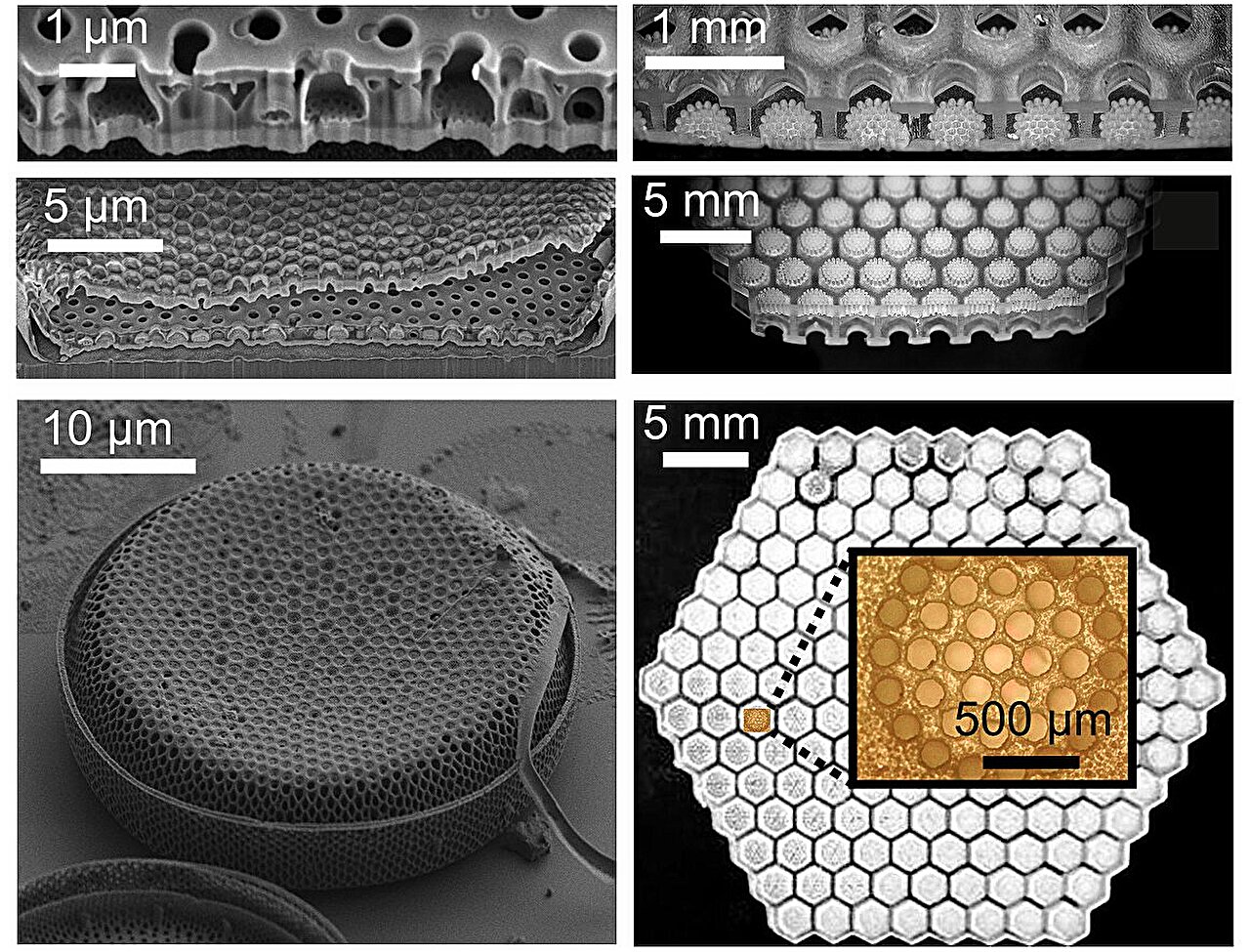
A diatom shell terahertz imaging experiment has confirmed an optical effect that researchers say could help improve future biomedical scanning and industrial inspection systems. Scientists from Skoltech, the Kurchatov Institute and institutes of the Russian Academy of Sciences built a 2,000-times enlarged copy of a microalga shell and tested it with terahertz waves, according to Skoltech.
The work, published in Light: Advanced Manufacturing, experimentally verified a previously predicted version of the Talbot effect in a structure modeled on a diatom shell. Skoltech said the finding points to possible new designs for compact optical parts that can focus and shape terahertz wavefronts.
What did the diatom shell terahertz imaging study show?
The researchers recreated the shell of a single-celled diatom alga, a tiny organism whose glass-like exterior has a complex pattern of pores and layers. In nature, that shell acts as a photonic structure, redistributing light that passes through it, according to the study.
The team used the enlarged model to observe how the shell-like geometry produces the Talbot effect in the terahertz range. The Talbot effect is a wave phenomenon in which a repeating structure creates self-images of its pattern at certain distances after light or another electromagnetic wave passes through it.
In living diatoms, the relevant interaction occurs with visible light. By scaling the structure up by a factor of 2,000, the researchers shifted the experiment into the terahertz band because the wavelength that interacts with a grating changes with the grating’s size, Skoltech said.
Previous terahertz studies of the Talbot effect had mainly used flat, simpler gratings, according to Skoltech. For a diatom-shell-like geometry, the effect had been predicted earlier by the same research group but had not been confirmed experimentally.
How the researchers built the enlarged shell
Making the model required a detailed reconstruction of the diatom shell’s three-dimensional shape. The team recorded the shell from many angles with scanning electron microscopes and atomic force microscopes, then used those images to rebuild the geometry.
The researchers manufactured the enlarged structure with LCD 3D printing, an additive manufacturing method. Skoltech said the printing process had to be adjusted carefully because the shell’s architecture includes a multilayered network of tiny openings.
Images released with the study show microscope views of the original shell alongside photographs of the enlarged model. The comparison underscores why the larger version was useful: it preserved the physical pattern while making the experiment easier to carry out at longer wavelengths.
Why terahertz waves matter for medical scans
Terahertz waves, also called T-waves, sit in a part of the electromagnetic spectrum that researchers are studying for biomedical visualization and nondestructive testing. Skoltech said T-waves are sensitive to water molecules in biological tissue and are safer for the body than X-rays and gamma rays.
That sensitivity could be useful in medical imaging, including possible skin cancer diagnostics, according to Skoltech. In industry, the same type of radiation can pass through dielectric materials and reveal internal defects without cutting into the material.
A barrier to wider use is the limited supply and high cost of terahertz-ready optical components, including mirrors, lenses and diffractive elements, Skoltech said. The study presents the enlarged diatom shell as a model for designing such components rather than as a finished medical device.
Julijana Cvjetinovic of Skoltech Photonics, a study co-author and grant project lead, said future work could use the diatom-inspired architecture while choosing lower-loss materials and adjusting the geometry for specific tasks. Dmitry Gorin, who leads the Biophotonics Laboratory at Skoltech Photonics and served as principal investigator, said the project also shows how scaling up an object can simplify an experiment while preserving the physical picture being tested.
This story draws on original reporting from Medical Xpress.