
Blood vessel formation mechanism shows how cells push, anchor and pull
University of Basel researchers tracked how endothelial cells connect hollow vessel segments, a finding that could aid organoid research.
By Tom Brennan · Health & Medicine Correspondent
3 min read
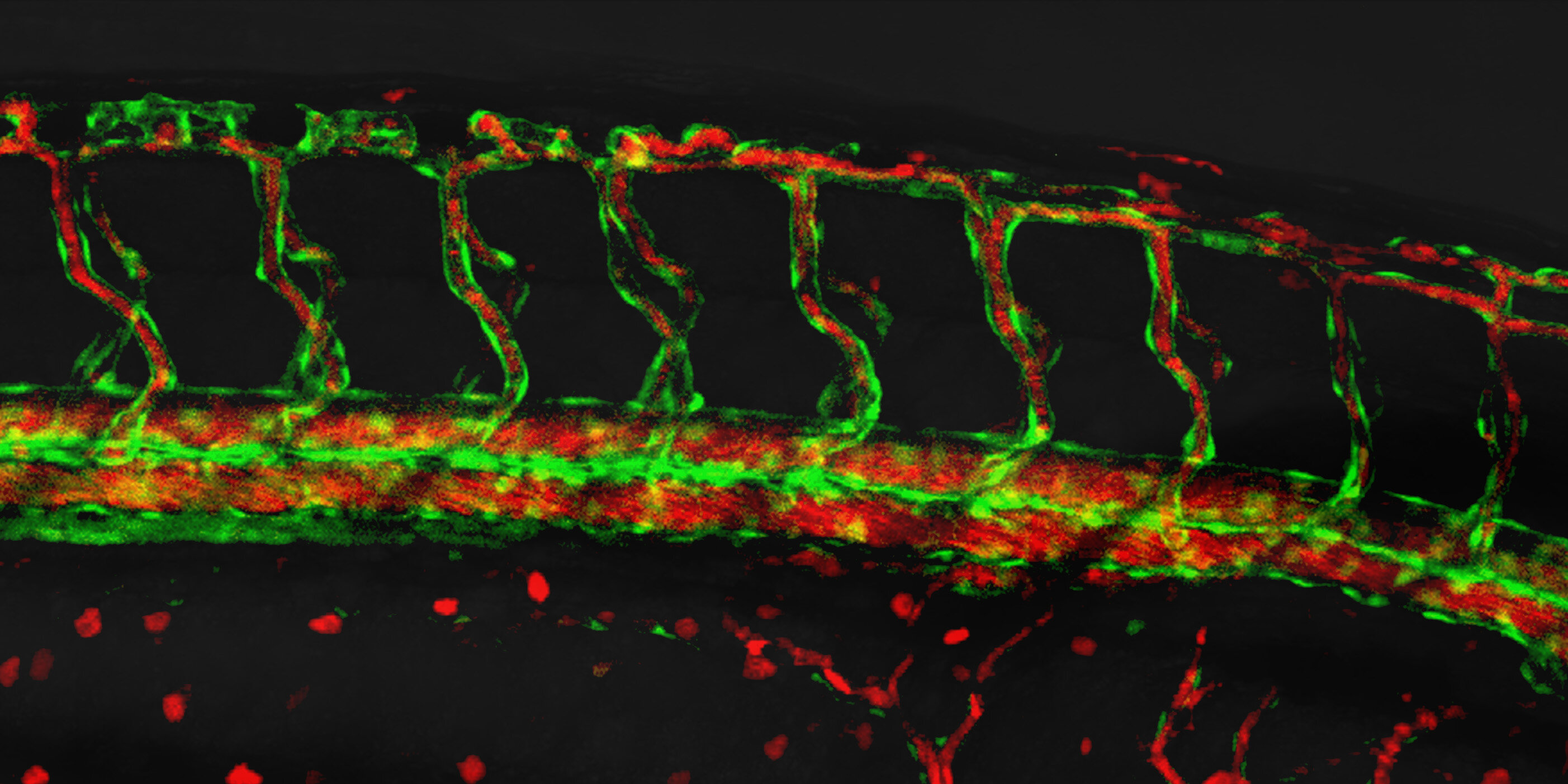
University of Basel researchers say they have identified a blood vessel formation mechanism that lets cells connect separate hollow channels into one continuous tube. The work helps explain how developing vessels become networks capable of carrying blood, oxygen and nutrients through the body.
The team, led by Professor Markus Affolter and Dr. Heinz-Georg Belting at the Biozentrum, used high-resolution live imaging in zebrafish to follow endothelial cells as vessels formed. The findings were published in eLife.
Blood vessels form from endothelial cells, which must build hollow tubes with an open internal space called a lumen. According to the University of Basel, scientists had only a partial picture of how these cells reshape themselves and coordinate movement while keeping the vessel wall intact.
How do cells build blood vessels?
The researchers report that endothelial cells use a repeated sequence of mechanical actions: one part of the cell pushes forward, attaches to a neighboring cell, and then pulling forces draw the rest of the cell along. That sequence lengthens the lumen and allows nearby vessel sections to join.
The process begins with a membrane extension at the front of an endothelial cell. The University of Basel described this extension as a junction-based lamellipodium, a structure the same research group had previously linked to cell rearrangement during vessel growth.
In the new study, the team says the protrusion generates forward force and then fastens to a neighboring cell at its tip. That attachment stabilizes the developing vessel connection before pulling forces move the rear part of the cell forward.
First author Dr. Ludovico Maggi compared the motion to an inchworm, saying the repeated push-and-pull cycle moves cells ahead while extending the lumen. As this happens, separate hollow vessel segments merge into channels with uninterrupted internal openings, according to the study.
What role does VE-cadherin play?
The researchers found that VE-cadherin, a molecule involved in endothelial cell junctions, has two roles in the process. Belting said it helps hold endothelial cells together while also taking part in cell movement.
That dual role is important because vessel-building cells face competing demands. They need firm junctions to preserve the vessel wall, but those same contact points must be flexible enough for cells to shift position and connect forming tubes.
Belting said the team was struck by the plasticity of the cells, which remained stable while also changing shape and position. The study links that behavior to coordinated cell junction activity and actomyosin dynamics, according to the eLife publication details.
Why the finding matters for organoids
The University of Basel said the findings could support future work on vascularized organoids, which are laboratory-grown tissues designed to more closely model natural organ development. Functional blood vessels are considered a key requirement for making such tissues more realistic for research and regenerative medicine.
Belting said blood vessels do more than supply tissues, because they develop alongside almost every organ. The researchers argue that understanding how vessels form also adds to the broader picture of how organs take shape.
The study was published as “Junctional and Actomyosin Dynamics Drive Endothelial Cell Rearrangements during Vascular Tube Formation” in eLife, with Maggi and colleagues listed as authors.
This story draws on original reporting from Medical Xpress.