
AI method could speed tissue checks during liver cancer surgery
Researchers reported an OCT imaging and AI approach that classified liver tissue within seconds in laboratory testing.
By Tom Brennan · Health & Medicine Correspondent
3 min read
Researchers have shown that an AI method paired with optical imaging can identify suspicious liver tissue within seconds, a step they say could shorten some liver cancer operations. The work, reported in Scientific Reports, is still at the laboratory stage and would need testing in operating rooms before clinical use.
According to Fraunhofer Austria Research GmbH, liver cancer surgery often includes frozen-section analysis of removed tissue so clinicians can check whether malignant tissue remains. Fraunhofer said that process takes place while the patient is still under general anesthesia, keeping staff and operating rooms occupied and adding time during which complications can occur.
Optical scans paired with anomaly detection
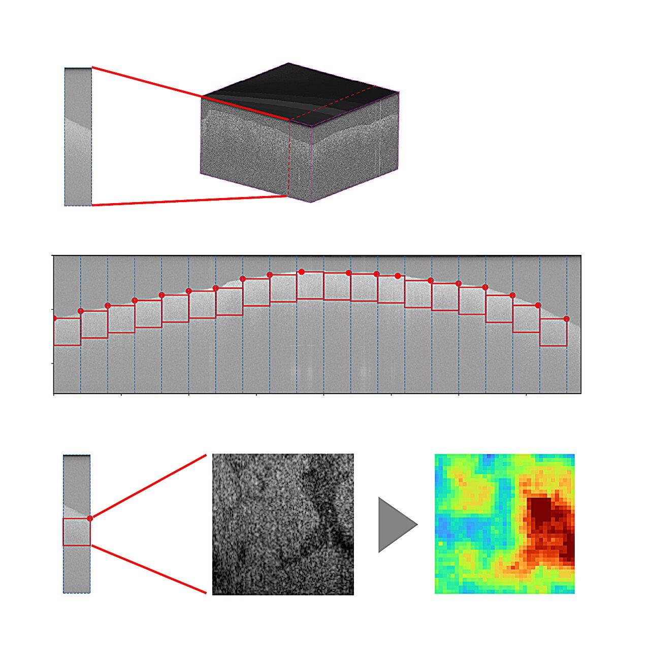
The research team tested optical coherence tomography, or OCT, a light-based imaging technique that Fraunhofer said is already used in ophthalmology to examine structures such as the optic nerve. OCT creates three-dimensional cross-sectional tissue images and, according to the researchers, can acquire those scans in seconds.
University Hospital RWTH Aachen proposed testing OCT for use during tumor removal, according to Fraunhofer. Working with the Fraunhofer Institute for Production Technology IPT, the team collected 173 OCT scans from 69 patients under laboratory conditions. Fraunhofer said the data included 88 scans of normal liver parenchyma and 85 scans covering different tumor types.
The team then applied anomaly detection, a machine-learning approach suited to situations where researchers have more examples of healthy tissue than diseased tissue, according to Fraunhofer Austria. Ulrich Krispel, an anomaly detection specialist at Fraunhofer Austria, said the model was trained only on normal liver scans and then used to flag tissue patterns that departed from that normal range.
In the published study, Krispel and colleagues reported a mean accuracy of 81% across the available data. Fraunhofer described the work as proof of concept for using anomaly detection as a decision-support tool in this setting.
Results still need surgical testing
Fraunhofer said the combined method produced classifications after only a few seconds of computation, identifying whether OCT scans showed normal liver parenchyma or tumor tissue. The team said standard histopathological examination would still be used to verify the results.
The reported accuracy differed by tumor type, according to the study. Fraunhofer said one tumor type was recognized with 94.3% accuracy, another with 84.5% accuracy and a third with 65.9% accuracy.
Iakovos Amygdalos of University Hospital RWTH Aachen, who Fraunhofer said first suggested exploring OCT for this use, said the approach could help create a faster intraoperative diagnostic tool for suspicious liver lesions. Fraunhofer said such a tool could reduce operation time, ease staff workload and make procedures less burdensome for patients if later studies support its use.
Caroline Girmen of Fraunhofer IPT said the project lays the groundwork for establishing OCT as an imaging method during liver surgery. According to Fraunhofer, the next research steps are to test the technology under real operating conditions and shrink the sensor system so it can fit into surgical workflows as a complement to histopathology.
The study, titled “Differentiating malignancy from liver parenchyma in Ex-Vivo OCT images using anomaly detection,” was published in Scientific Reports by Krispel and colleagues.
This story draws on original reporting from Medical Xpress.