
Cerebral schistosomiasis diagnosed in Philippine woman after antibody test
Doctors linked a Philippine rice farmer’s focal seizure to Schistosoma japonicum after MRI, blood-serum and stool testing.
By Maya Lindqvist · Senior Technology Correspondent
2 min read
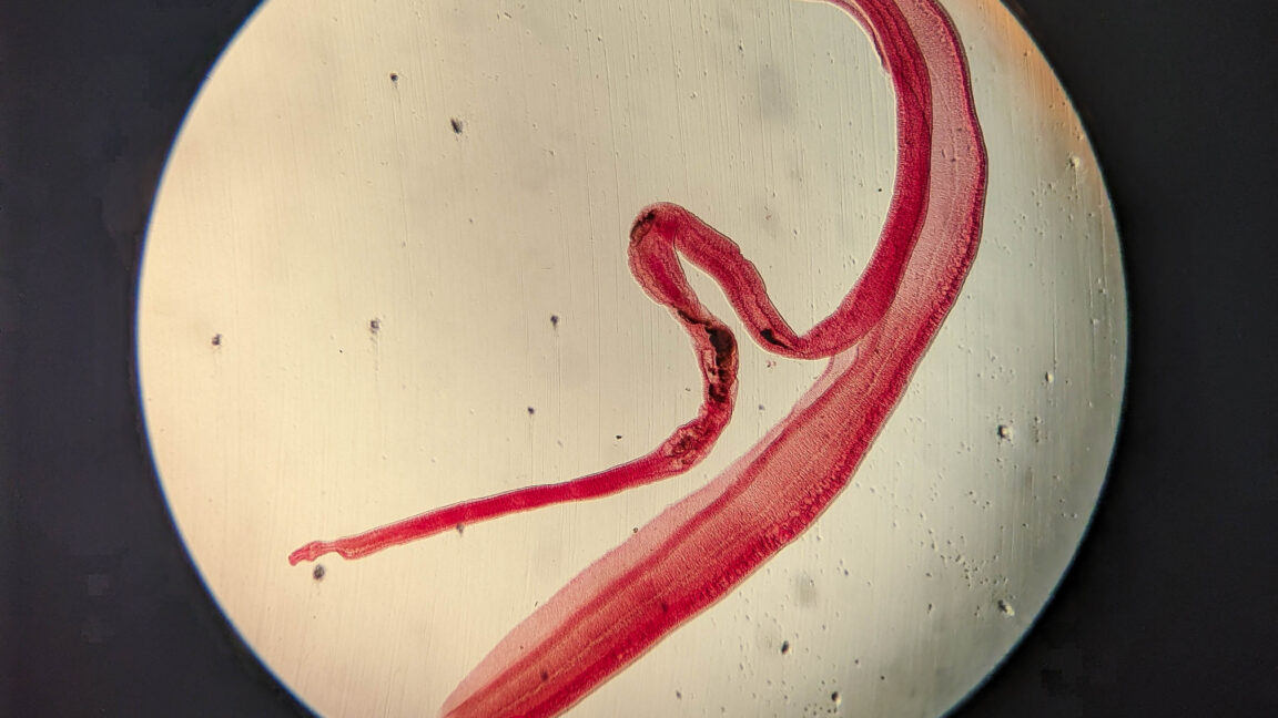
A case report has documented cerebral schistosomiasis Philippines in a rice farmer whose sudden shaking of her right arm and leg was linked to a Schistosoma japonicum infection. The tail-like forms seen during testing were an antibody reaction around laboratory reference eggs, not eggs in the patient growing tails.
According to the report described by Ars Technica, the woman had about one minute of uncontrolled shaking on her right side. She had not reported headache, confusion or weakness before the episode, and an initial physical examination did not find an abnormality.
An MRI then showed a cluster of bright nodules surrounded by fluid in the left frontal lobe. Doctors suspected that the nodules represented parasite eggs and began tests for schistosomiasis, the report said.
How was cerebral schistosomiasis diagnosed in this case?
Doctors used a circumoval precipitin test, which combines a patient’s serum with reference S. japonicum eggs. If the serum contains antibodies directed at the parasite, the antibodies bind material on or released by the eggs, creating a tail-like precipitate outside the egg.
The test produced that result for this patient, supporting an S. japonicum infection. Stool testing also found 190 S. japonicum eggs per gram of feces. With those findings and the MRI, doctors diagnosed cerebral schistosomiasis causing a focal seizure, according to the case report.
S. japonicum is a blood fluke in the Schistosoma group and is the most common such fluke in the Philippines, Ars Technica reported. The report describes brain involvement as an uncommon complication in which eggs reach the central nervous system; eggs lodged in the brain are associated with seizures or focal neurological problems.
What treatment and outcome did doctors report?
The patient received an antiparasitic medicine, an anti-inflammatory medicine and an antiseizure medicine. Three months later, she reported no further seizures, and a follow-up MRI showed that the eggs were no longer visible, according to the report.
The diagnosis is distinct from a widely reported Australian case in 2023. In that case, a live Ophidascaris robertsi roundworm was surgically removed from a woman’s brain; it involved a different parasite, country and diagnostic course, the BBC reported.
This story draws on original reporting from Ars Technica.