
Cryo-EM study maps bacterial protein gate for folded cargo
ISTA researchers used cryo-electron microscopy to show how the Tat system may move large folded proteins across cell membranes.
By Priya Raghavan · Science Reporter
3 min read
Researchers at the Institute of Science and Technology Austria have mapped new structural details of a bacterial membrane machine that moves large, already folded proteins without tearing the membrane apart. The work matters because the system, known as twin-arginine translocase, or Tat, is found in bacteria and chloroplasts but not in humans, making it a possible target for antimicrobial strategies.
The findings by Leonid Sazanov and postdoctoral researcher Ziyu Zhao were published in Molecular Cell, according to ISTA. The study focused on the Tat system in E. coli, where scientists had already identified three components called TatA, TatB and TatC, but had not resolved how the assembled complex handles bulky protein cargo.
Cells contain membrane-bound compartments, and many proteins must cross those barriers to work in the right place. ISTA said many cells use the Sec system for this task, pulling proteins through membranes while they are unfolded, a process that is easier than moving a fully folded protein.
The Tat system addresses the harder case, according to the researchers. It transports proteins that have already folded into their working shape, which makes them too large to pass through a narrow channel in the same way as an unfolded chain.
How the structure was captured
Zhao isolated the Tat complex directly from living E. coli cells, ISTA said. The team reported that the complex was unstable until the Tat components and the cargo protein were produced together in the bacteria, which made the assembly stable enough for imaging.
The researchers placed the Tat complex with bound cargo on electron microscopy grids and rapidly cooled it in cryogen. According to ISTA, the fast freezing preserved the sample in a glasslike state, avoiding ice crystals that could damage fine structural features.
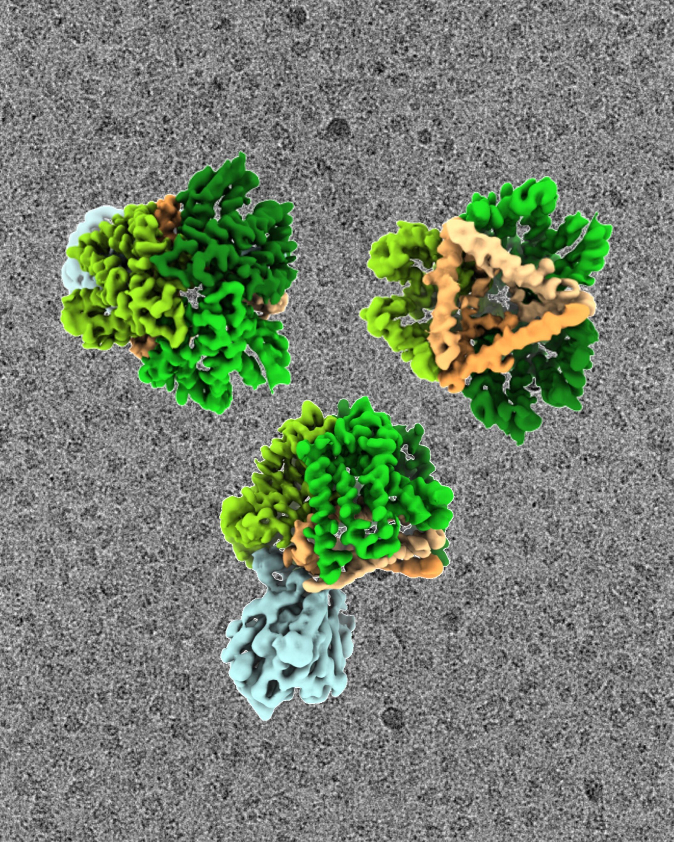
The team then used cryo-electron microscopy, a method capable of resolving protein structures at about 2 to 3 angstroms, ISTA said. About 10,000 images were analyzed to build the reconstruction, according to the image information released with the study.
A bowl-shaped molecular machine
The three-dimensional reconstruction showed a complex made from three TatB/C units that sit in the membrane, according to ISTA. Sazanov described the structure as resembling an open bowl with a thin base.
The researchers found that the cargo binds to the bowl-shaped complex at two sites on the TatB/C units, ISTA said. One site recognizes the cargo’s signal peptide and holds it in place, while a second site appears to check whether the protein is correctly folded before transport proceeds.
The reconstruction also suggested that the base of the complex may contain a pore that can open and close, according to Sazanov and Zhao. The researchers propose that this pore could act as a gate, opening after the cargo has docked and passed the folding check, allowing the protein to cross the membrane intact.
ISTA said the precise steps of that gating process are still unresolved and will be a focus of Zhao’s future research. The published paper is titled “Structure of E. coli twin-arginine translocase (Tat) complex with bound cargo,” with DOI 10.1016/j.molcel.2026.05.026.
The system’s absence from humans gives the work possible medical relevance, according to ISTA. In bacteria, Tat is tied to metabolism and virulence, so a clearer view of its components could help efforts to design treatments that disrupt the process in harmful bacteria.
This story draws on original reporting from Phys.org.