
Penn researchers identify retinal cell stages that could improve transplants
A mouse study points to three photoreceptor precursor states that may help scientists choose better cells for future vision-restoring therapies.
By Tom Brennan · Health & Medicine Correspondent
3 min read
Researchers at the Perelman School of Medicine at the University of Pennsylvania have identified three developmental states of retinal photoreceptor cells, a finding they say could help improve future cell transplants for blinding eye diseases. The work matters because current photoreceptor transplants have shown limited success, with few donor cells connecting well enough in the retina to restore vision.
The study, published June 12 in Frontiers in Cell and Developmental Biology, examined photoreceptor development in mice. Photoreceptors are the light-sensing neurons in the retina, the tissue at the back of the eye that converts light into signals the brain can interpret.
The Penn team reported that developing photoreceptor cells can be grouped into early, middle and late states. The researchers said those distinctions may help identify which cells are most likely to survive after transplant and become part of the eye’s circuitry.
Retinal diseases affect millions of people worldwide and are a leading cause of blindness, according to the university. Some are inherited, while others, including age-related macular degeneration, do not have a known genetic cause. Existing treatments focus mainly on slowing or stopping vision loss, while cell-based approaches aim to replace cells that have been lost or damaged, the researchers said.
Katherine Uyhazi, the study’s principal investigator and an assistant professor of ophthalmology at Penn, said the team plans to test the cell groups separately. “We plan to isolate and transplant each subgroup individually, in the hopes that transplanting a more pure cell population will improve future cell-based therapies to improve vision in late-stage blinding conditions,” Uyhazi said.
Retinal development in waves
The study builds on the idea that retinal cells do not all mature at the same pace. Uyhazi said retinal development occurs in waves, leaving a mixture of cell stages present at the same chronological point during development.
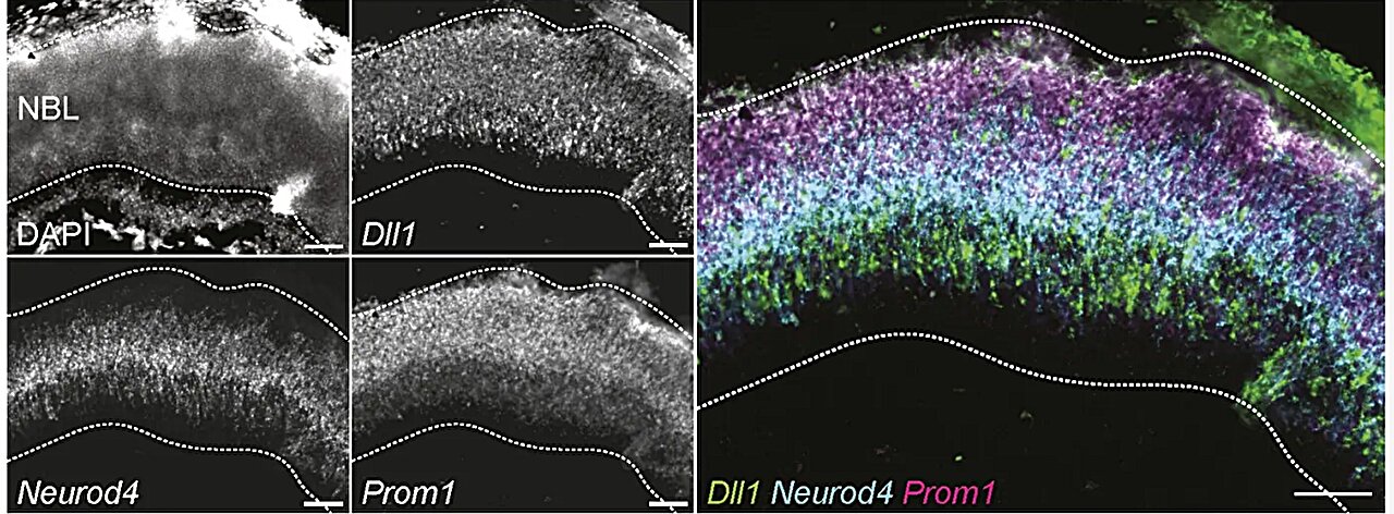
Joseph Yano, a Ph.D. candidate in Uyhazi’s laboratory and the study’s first author, used single-cell RNA sequencing to examine gene activity in individual cells, according to Penn. That analysis allowed the researchers to separate developing photoreceptors into the three states they described.
The team also found evidence that similar cell populations may exist in human retinal organoids. Those organoids are three-dimensional structures grown in the laboratory to model parts of the human eye, according to the university.
Next tests in mice
Uyhazi’s group is now developing ways to isolate and transplant each cell state into the retina, Penn said. The researchers said early-stage cells resemble stem cells more closely and may be better able to survive transplantation, while late-stage cells are more like mature retinal cells capable of responding to light.
The group plans further mouse experiments to determine whether one developmental stage offers the best balance for retinal repair. Penn said the goal is to find a cell population that can survive transplantation and connect with the eye more effectively than mixed or less-defined donor cells.
The paper, “Lineage tracing reveals photoreceptor precursor cell subpopulations that contribute to murine retinogenesis,” was authored by Joseph J. Yano and colleagues and published in Frontiers in Cell and Developmental Biology.
This story draws on original reporting from Medical Xpress.