
Mouse virus study offers Parkinson’s model without toxins
Texas A&M researchers say a mouse virus produced Parkinson’s-like brain damage and movement problems, giving scientists a new way to study possible viral triggers.
By Tom Brennan · Health & Medicine Correspondent
3 min read
A Texas A&M University research team reports that a natural mouse virus can produce Parkinson’s-like brain injury and movement problems in mice. The finding matters because it gives researchers a possible disease model that does not rely on genetic engineering or toxic chemical exposure, according to Texas A&M.
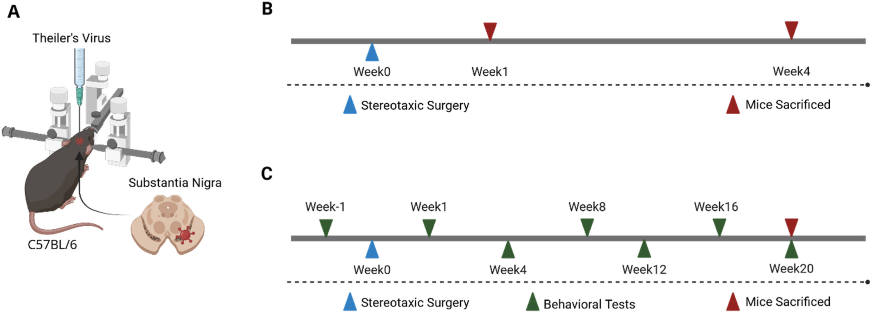
The study, published in Brain, Behavior, & Immunity - Health, tested Theiler’s murine encephalomyelitis virus, or TMEV, as an infectious trigger in a pilot mouse model of Parkinson’s disease. TMEV is a natural pathogen in mice, according to the university.
Parkinson’s disease affects more than 10 million people worldwide, Texas A&M said. The disease damages dopamine-producing cells in the brain; dopamine is needed for smooth movement, and loss of those cells is tied to tremors, stiffness, walking and balance problems, as well as mental or emotional distress.
Why researchers tested a virus
Animal models are central to Parkinson’s research, but Texas A&M said many existing models depend on gene changes or toxicants. Candice Brinkmeyer-Langford, a neurodegenerative disease expert at the Texas A&M University School of Public Health, said toxic-exposure models help researchers study Parkinson’s but cannot capture every way the disease may begin or progress in people.
Researchers have long suspected that infections could help set off Parkinson’s through inflammation in the brain, according to Texas A&M. The university said scientists also consider genetics and environmental exposures part of the disease’s possible origins, which remain unknown.
Brinkmeyer-Langford said viruses can lead to different illnesses depending on a person’s genetics. She cited Epstein-Barr virus, which causes mononucleosis and may also contribute to cancer or multiple sclerosis, and SARS-CoV-2, which can affect the heart and brain as well as the lungs.
What the mouse tests found
The researchers examined whether TMEV infection damaged dopamine-producing brain cells and caused movement problems similar to those seen in Parkinson’s, according to Texas A&M. One week after infection, they confirmed that the virus had reached dopamine-producing cells; one month after infection, those cells had been destroyed at the infection site.
The team compared 13 infected mice with 14 healthy controls after giving them a drug that mimics dopamine, Texas A&M said. The resulting movement pattern supported the conclusion that the infected mice had lost dopamine neurons.
The study also used a pole test to measure speed and coordination. Texas A&M said infected mice took longer than healthy controls to complete the test, and the slower performance was still present at week 20, when the study ended.
Researchers also used a specialized treadmill to assess gait, balance and motor function across more than 100 walking-related measures, according to Texas A&M. The university said those tests showed physical weakness after dopamine-producing cells were lost, indicating brain damage similar to that associated with Parkinson’s patients.
Next research steps
Brinkmeyer-Langford said future work will compare the TMEV model with older standard animal models used in Parkinson’s research. Texas A&M said the team also plans to search for early warning signs and biological markers and to study how immune responses to viral infection alter the brain.
The paper is titled “Theiler’s murine encephalomyelitis virus as the infectious agent for a virally induced mouse model of Parkinson’s disease.” Its DOI is 10.1016/j.bbih.2026.101230.
This story draws on original reporting from Medical Xpress.