
Mouse study links cerebellar cell coatings to social behavior
Kanazawa University researchers report that disrupting perineuronal nets in the cerebellum altered social behavior in autism-related mouse models.
By Priya Raghavan · Science Reporter
3 min read
Kanazawa University researchers have reported a cerebellar mechanism that may help explain changes in social behavior seen in autism-related mouse models. The study, published in Translational Psychiatry, points to perineuronal nets, extracellular structures that surround some neurons, as regulators of cerebellum-linked circuits involved in social interaction.
Autism spectrum disorder is marked mainly by differences in social communication and interaction, according to Kanazawa University. The university said growing evidence suggests ASD involves changes across connected brain circuits rather than a problem confined to one brain area.
The cerebellum has long been associated with movement control, but Kanazawa University said recent research has tied it to cognition, emotion and social behavior. The new work examined how changes in cerebellar circuits could affect social behavior in mouse models linked to ASD risk.
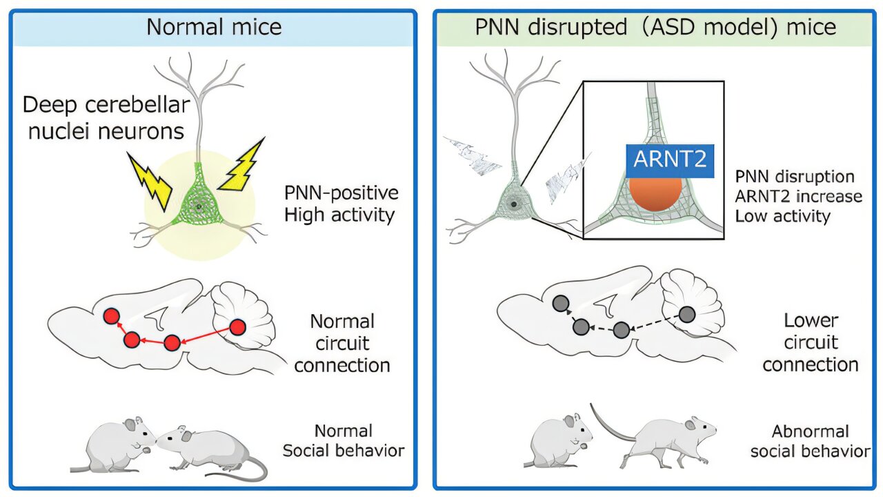
The research team studied two different mouse models, according to Kanazawa University: mice exposed prenatally to valproic acid, an environmental risk model, and mice carrying a mutation in Chd8, a gene associated with ASD risk. The group looked for brain changes that appeared in both models.
In both sets of mice, the researchers found fewer perineuronal nets around neurons in the deep cerebellar nuclei, a major output area of the cerebellum, Kanazawa University said. Perineuronal nets help maintain neuronal excitability, shape synaptic signaling and support circuit maturation, according to the university.
To test whether those structures affected behavior, the team used an enzyme-based method to break down perineuronal nets in the cerebellar nuclei, Kanazawa University said. Mice with disrupted nets showed reduced social interaction and less interest in unfamiliar mice, findings the researchers interpreted as evidence that intact nets in this cerebellar region are needed for typical social behavior in mice.
The study also examined circuit activity. Kanazawa University said social stimuli usually activated neurons in the cerebellar nuclei and that the signal extended to connected regions including the midbrain and thalamus.
That pattern changed when perineuronal nets were disrupted, according to the university. The affected mice showed weak activation in cerebellar nuclei neurons and lower activity across circuits connected to the cerebellum, suggesting that local structural changes in the cerebellum can alter wider networks tied to social behavior.
The researchers also identified increased expression of ARNT2, a transcription factor, in neurons that lacked perineuronal nets, Kanazawa University said. The university said ARNT2 controls gene expression related to neuronal activity, and higher ARNT2 levels appeared to make neurons less responsive.
Suppressing ARNT2 restored neuronal activity and social behavior in the experiments, according to Kanazawa University. The researchers described ARNT2 as a molecular link between perineuronal net loss and circuit dysfunction.
Kanazawa University said the findings broaden the focus of ASD-related circuit research, which has often centered on the cerebral cortex or synaptic function. The study highlights extracellular matrix structures in the cerebellum as contributors to brainwide activity patterns that influence social behavior in mice.
The university said future work will examine whether similar mechanisms operate in humans and whether changing cerebellar circuit activity can affect social behavior. The paper was authored by Kyota Fujita and colleagues and published under the title “Perineuronal nets in cerebellar nuclei neurons orchestrate social behaviour via regulation of neuronal activity in circuits innervated by the cerebellum.”
This story draws on original reporting from Medical Xpress.