
Immune cell niche tied to joint tissue overgrowth in rheumatoid arthritis
Researchers linked SPP1-high macrophages and fibrin-rich joint niches to abnormal synovial growth in rheumatoid arthritis.
By Tom Brennan · Health & Medicine Correspondent
3 min read
Researchers at Hospital for Special Surgery say they have identified an immune-cell niche that helps drive abnormal tissue growth in rheumatoid arthritis. The finding matters because joint damage in the disease is tied not only to inflammation, but also to expansion of the synovial tissue that can invade cartilage and bone.
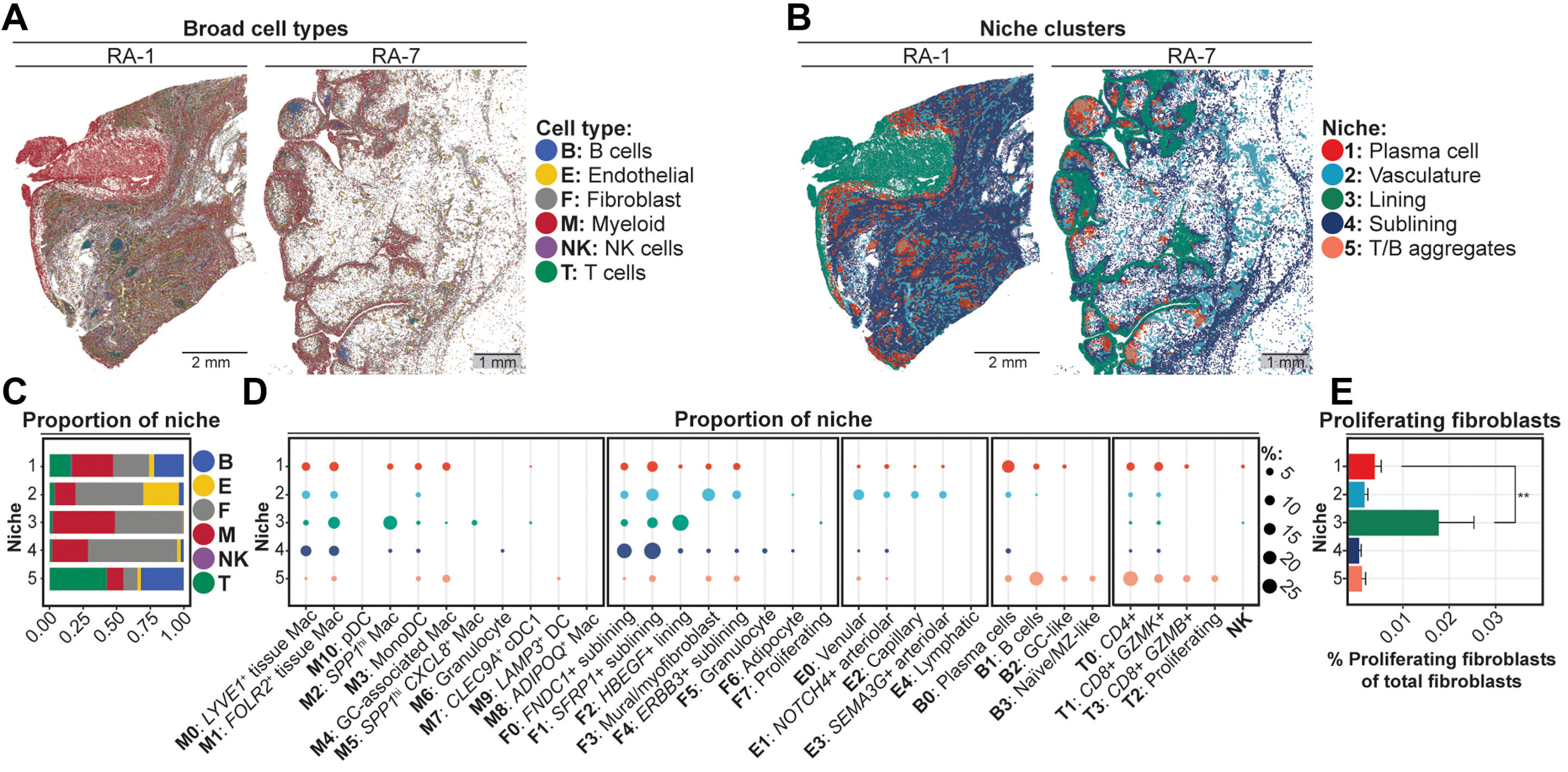
The study, published in Science Translational Medicine, focused on a subset of macrophages known as SPP1-high macrophages. According to the HSS team, these immune cells appear to work with fibroblasts and a temporary protein framework made of fibrin to promote growth and remodeling inside affected joints.
How the joint lining changes
Rheumatoid arthritis is a chronic autoimmune disease in which inflammation affects the synovium, the tissue that lines joints. Hospital for Special Surgery said that over time the disease can cause pain, swelling and damage to cartilage and bone.
A key problem in rheumatoid arthritis is pannus, an invasive overgrowth of synovial tissue. Current treatments often aim to reduce inflammation, while the processes that cause synovial tissue to expand have been less clear, according to the researchers.
Using spatial transcriptomics on human tissue samples, the HSS team found that SPP1-high macrophages gather in areas of the synovium that are rich in fibrin. Fibrin is best known for its role in clotting and wound repair, and the researchers said it may serve as a short-term scaffold for tissue formation in the rheumatoid joint.
Macrophages, fibrin and fibroblasts
The study reported that SPP1-high macrophages in these fibrin-rich niches break down fibrin through enzymes and cellular uptake. The same niches were associated with nearby fibroblasts that were primed to multiply and deposit matrix, according to the paper.
Hospital for Special Surgery said the results point to a coordinated process involving immune cells, structural cells and the extracellular matrix. Laura Donlin, senior author and a scientist at the HSS Research Institute, said the work supports a view of rheumatoid arthritis in which faulty tissue repair contributes to disease progression alongside inflammation.
The researchers distinguished the process from classic fibrosis. Although the macrophages resemble cells implicated in fibrotic conditions of organs such as the lung and liver, the rheumatoid arthritis samples did not show the dense collagen accumulation typical of fibrosis, according to the study.
Instead, the authors described the pattern as pro-generative tissue remodeling. In their interpretation, the joint environment shows features more similar to wound healing that has become misdirected.
Possible treatment implications
The study suggests that SPP1-high macrophages and their signaling pathways could become targets for future rheumatoid arthritis treatment strategies. The authors also pointed to IL-6 signaling as one factor that helps sustain these cells.
That finding may help explain why IL-6-targeting therapies, which are already used in rheumatoid arthritis, can be effective for some patients, according to the HSS researchers. Donlin said the work points to disease-driving pathways beyond inflammation that could help guide more precise treatment approaches.
The authors said similar immune-cell populations and fibrin-linked remodeling have been implicated in interstitial lung disease, lupus, cancer and traumatic injury. They said that raises the possibility that the pathway has a broader role in human disease, though the study centered on rheumatoid arthritis synovium.
The paper was authored by Ian Mantel and colleagues and is titled “SPP1hi macrophages in fibrin niches promote hyperplastic tissue remodeling in rheumatoid arthritis synovium.”
This story draws on original reporting from Medical Xpress.