
Corneal mechanics imaging prototype records nine eye points at once
ICTER researchers built an OCT prototype that records nine corneal points at once, aiming to spot localized weakening earlier.
By Priya Raghavan · Science Reporter
3 min read
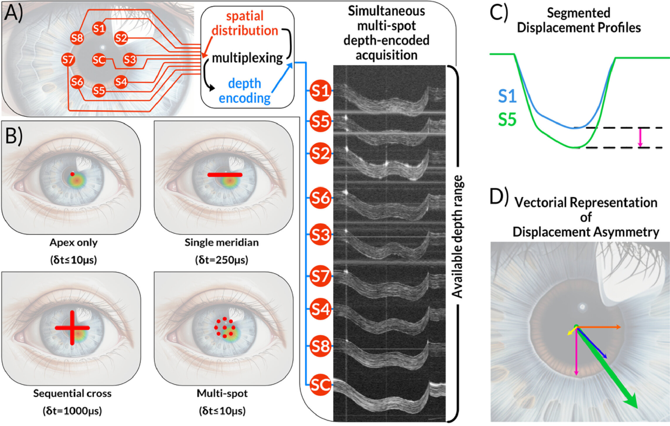
A corneal mechanics imaging system developed by researchers at the International Centre for Translational Eye Research can record the eye’s response to an air pulse at nine locations at the same time. The prototype matters because localized corneal weakening, including in keratoconus, may be missed when instruments measure only an overall response or a single cross-section.
The work, carried out at ICTER within the Institute of Physical Chemistry of the Polish Academy of Sciences, was described in Biomedical Optics Express. The study’s authors include Karol Karnowski, Jadwiga Milkiewicz, Onur Cetinkaya, Angela Pachacz, Andrea Curatolo, Kamil Liżewski, Dawid Borycki and Maciej Wojtkowski, with collaborators from institutions in Poland, the United Kingdom, Spain and the United States.
How does the corneal mechanics imaging method work?
The prototype uses optical coherence tomography, or OCT, an imaging method that uses light to produce high-resolution views of eye tissue. In this system, several light beams examine the cornea at once while a short air pulse causes the surface to deform.
The system measures one central point and eight surrounding points. ICTER researchers said the signals are separated by depth encoding, allowing the device to capture multiple locations in a single OCT acquisition rather than scanning them one after another.
That timing is central to the method. The study reports that the corneal motion is recorded every 10 microseconds, equal to 100,000 measurements per second across all nine points, while the deformation event lasts about 20 milliseconds.
According to the researchers, slower sampling produced substantial errors when estimating corneal asymmetry. When the data were degraded to 250 microseconds, the error in the magnitude of the asymmetry vector reached about 20%; at 1 millisecond, it approached 70%. Directional error rose to 17.5 degrees at 250 microseconds and 47.5 degrees at 1 millisecond.
Why keratoconus is a target
Keratoconus is a progressive condition in which the cornea thins and bulges outward, affecting vision. The Polish Academy of Sciences said mechanical changes may appear before clear changes in corneal shape, making biomechanical measurement a possible route to earlier detection.
Current eye instruments commonly measure corneal thickness, curvature and shape, and some assess the cornea’s response to a noncontact air puff. ICTER’s team focused on a limitation of those approaches: weakening may occur in a local region rather than evenly across the cornea.
To summarize the nine-point data, the researchers introduced an “asymmetry vector.” Its magnitude reflects the size of the biomechanical imbalance, while its direction points toward the region with the strongest deformation.
In patients with keratoconus, the study found that the vector pointed toward the affected corneal area, corresponding to regions that were thinner, steeper and had abnormal posterior elevation. The authors said the measurement complements tomography and topography because it assesses mechanical behavior rather than anatomy alone.
What the prototype still needs
The researchers also reported a previously unobserved “dual-indentation response,” in which the cornea showed two deformation phases after one air pulse. They observed it after widening the air nozzle from 1.5 millimeters to 3.7 millimeters, which broadened the pulse and cut peak force by about half while keeping the pulse duration at 8 milliseconds.
The study was a technical proof of concept, not a clinical validation. The researchers said a separate clinical dataset has been collected from healthy volunteers and patients with early and mild keratoconus, with results planned for a future publication.
The current system also has practical limits. ICTER said analysis now requires manual segmentation of the anterior corneal surface in OCT images, and the team is working on automated processing, including machine learning-based methods. Future versions may also add pupil monitoring, alignment feedback and eye tracking to reduce errors from movement, blinking or poor positioning.
This story draws on original reporting from Medical Xpress.