
Capillary cells kept signaling roles for weeks in mouse study
Yale and UCLA researchers found stable calcium-signaling networks in skin capillaries, with breakdowns tied to leakier vessels and altered blood flow.
By Priya Raghavan · Science Reporter
3 min read
Endothelial cells in mouse skin capillaries held distinct calcium-signaling roles for at least two weeks, according to researchers at Yale School of Medicine and UCLA. The finding, reported in the Proceedings of the National Academy of Sciences, suggests capillary communication is more organized and stable than previously shown in living mammals.
The work focused on endothelial cells, which line blood vessels and use calcium pulses as part of the signaling that helps regulate vessel behavior. Scientists have long known these signals occur, but the Yale-UCLA team reported that the same cells tended to remain either active or quiet across repeated observations.
The study was conducted in the laboratory of Valentina Greco at Yale, with close collaboration from the UCLA laboratories of Julia Mack and Chen Yuan Kam. Anush Swaminathan, an MD-Ph.D. student at Yale, was the paper’s first author, according to Yale University.
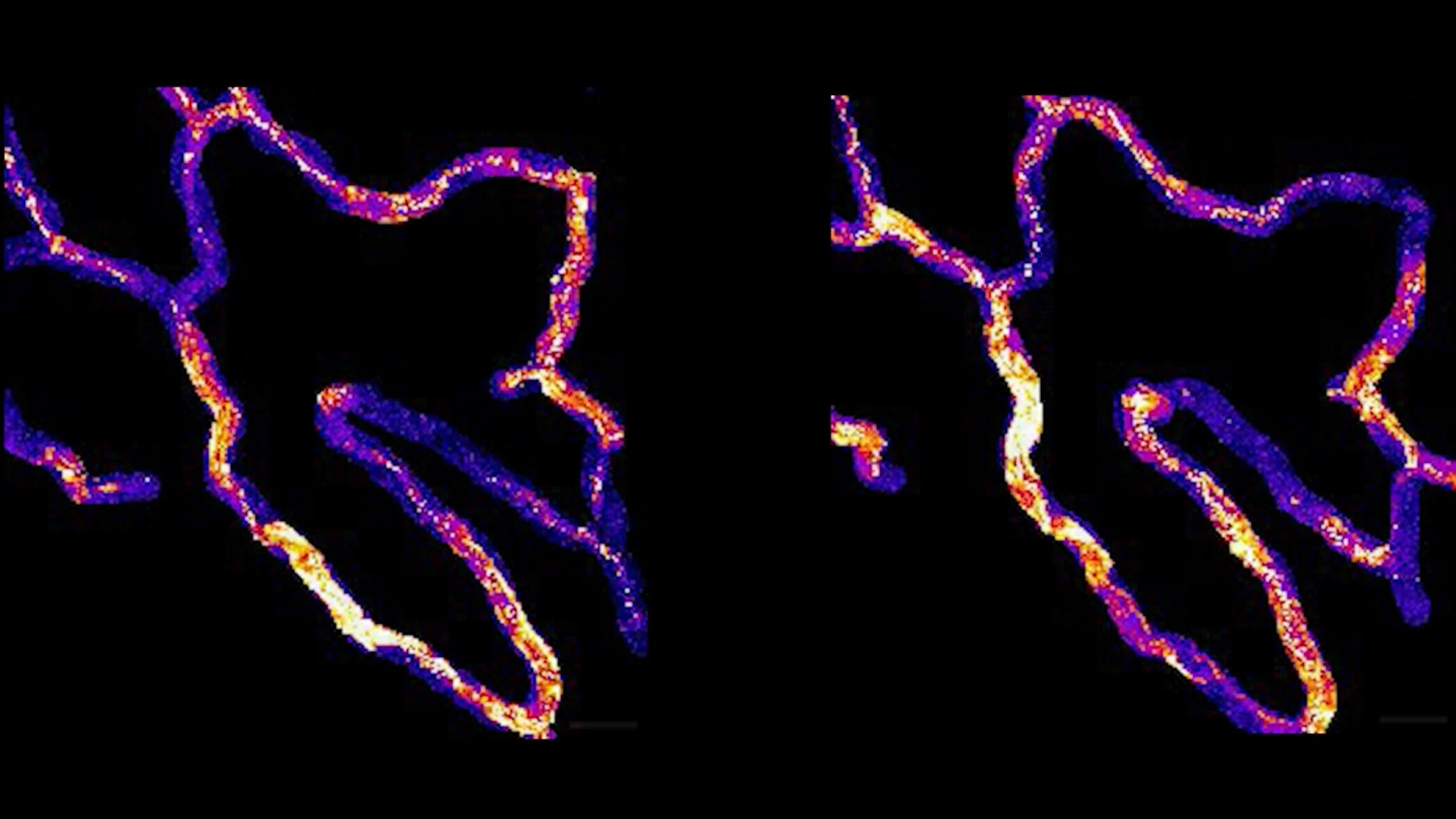
Tracking the same capillary cells
The researchers used genetically engineered mice whose endothelial cells glowed green when calcium was active. That allowed the team to watch signaling in living tissue without invasive procedures, Yale said.
Across thousands of cells in multiple mice, about half of the endothelial cells in a given region were firing calcium signals at a given time, while the rest were inactive, according to the study. When the researchers returned to the same animals 24 hours later, they found the same active cells signaling again. The pattern persisted when they checked again two weeks later.
Mack said the result echoed earlier work in cultured endothelial cells under flow, where a similar share of cells showed calcium activity. Kam said the findings build on evidence that adult endothelial cells keep stable positions in blood vessels, adding that the cells also appear to keep stable signaling identities.
The researchers also found variation inside that stable pattern. Individual cells changed the frequency and length of their calcium pulses from day to day, but the overall network maintained a consistent statistical profile, according to Yale.
What happened when communication failed
To test how the network stays coordinated, the team studied Connexin 43, or Cx43, a protein that forms channels for communication between neighboring cells. Yale said Cx43 is the most abundant connexin in skin endothelial cells.
When the researchers deleted Cx43 from endothelial cells, calcium signaling increased rather than decreased. Swaminathan said prior literature had suggested that removing gap junction channels would reduce communication between cells, but the team observed broader and longer calcium activity across the tissue.
Some cells fired continuously for 17 minutes, a pattern the researchers did not observe in typical mice, according to Yale. Over two weeks, more cells joined the persistently active group. The study also found increased capillary blood flow and leakier vessels, with molecules escaping from the bloodstream into surrounding tissue.
The team then screened a small group of drugs that target calcium channels. Nifedipine, a high blood pressure drug, stood out: when applied to a mouse paw, it restored calcium signaling, returned flow rates to baseline and repaired barrier function, according to Yale.
Yale said nifedipine acts on L-type voltage-gated calcium channels, which are not expressed in endothelial cells. Those channels are found in pericytes, support cells wrapped around capillaries, and possibly in upstream smooth muscle cells. The researchers reported that calming those neighboring cells appeared to affect endothelial behavior indirectly.
Unanswered questions
Swaminathan said preliminary work in younger developing mice also showed a conserved signaling network, suggesting the cell identities may form before adulthood. The researchers also found that individual calcium events in skin did not correlate with local blood flow changes, unlike findings reported in brain capillaries.
The study leaves open what single calcium pulses do in normal skin physiology. For now, the authors report that skin capillary endothelial cells form a stable signaling network over space and time, and that disruption of the network can coincide with impaired vascular function.
This story draws on original reporting from Medical Xpress.