
AI method produces pathology stains despite imperfect slide alignment
HKUST researchers say a generative AI framework can create virtual tissue stains that closely resemble chemical stains while preserving biopsy material.
By Tom Brennan · Health & Medicine Correspondent
3 min read
Researchers at the Hong Kong University of Science and Technology have developed a generative AI system designed to create virtual pathology stains even when training images do not line up precisely. The work matters because tissue staining is central to cancer diagnosis and research, but repeated chemical staining can take time and use up limited biopsy material.
The study, published in Nature Communications, describes a framework called Decoupled Generation and Registration, or DGR. HKUST said the method was built to address a common weakness in virtual staining systems: their reliance on image pairs that are assumed to be nearly perfectly aligned.
Pathology slides often fail that assumption, according to the research team. Tissue can deform during sectioning, staining, scanning, mounting, folding or damage, leaving the same structures slightly shifted between images. In AI training, that mismatch can make a correctly generated nucleus or tissue feature appear wrong because the reference image places it elsewhere.
How the framework works
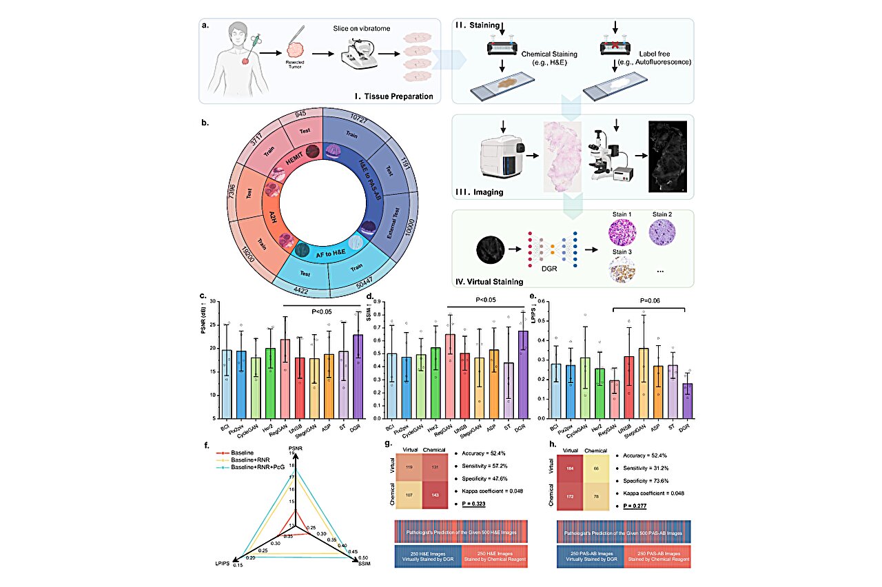
Virtual staining uses AI to convert one kind of tissue image into another, such as turning label-free autofluorescence images into H&E-like images or converting H&E images into PAS-AB-like special stains. HKUST said these digital stains could reduce the need for repeated chemical processing and add extra image channels for diagnosis, research analysis and multimodal models.
DGR separates two jobs that earlier approaches often combine. The generative part learns how one stain should look when transformed into another, while a registration component accounts for spatial shifts caused by tissue deformation, according to the study.
The team tested DGR on five datasets and four stain-related tasks. Those tasks included virtual H&E staining from label-free autofluorescence images, H&E-to-PAS-AB stain translation, H&E-to-multiplex immunohistochemistry conversion and H&E stain normalization.
Compared with other virtual staining models described by the researchers as state of the art, DGR showed stronger overall results in image quality and structural fidelity across the tasks. The study was led by Chen Hao of HKUST’s Department of Computer Science and Engineering, with Terence Wong of the Department of Chemical and Biological Engineering and collaborators from Southern Medical University, the Chinese University of Hong Kong and other partners.
Pathologists tested the images
To judge visual quality, the researchers asked experienced pathologists to review virtual and chemical stains without being told which was which. According to HKUST, the pathologists evaluated 500 H&E-stained images and 500 PAS-AB-stained images.
The pathologists distinguished DGR-generated stains from real chemical stains with about 52% accuracy, close to chance. The researchers said that result indicates the virtual and chemical stains were difficult to tell apart in that evaluation.
The team also tested whether the AI-generated stains could help other pathology AI models. When DGR-generated virtual multiplex immunohistochemistry images were paired with H&E images, model performance improved on colorectal polyp classification and gastric cancer tissue classification tasks, according to the study.
Chen, the paper’s corresponding author, said the work addresses a barrier to using virtual staining in clinical workflows by allowing high-quality results from imperfectly aligned pathology images. HKUST identified Ma Jiabo and Li Wenqiang, both Ph.D. students in Chen’s group, as co-first authors.
This story draws on original reporting from Medical Xpress.