
Second complete fruit fly brain map includes male nerve cord
A new male fruit fly connectome maps its brain and nerve cord, allowing scientists to compare neural wiring across sexes.
By Maya Lindqvist · Senior Technology Correspondent
3 min read
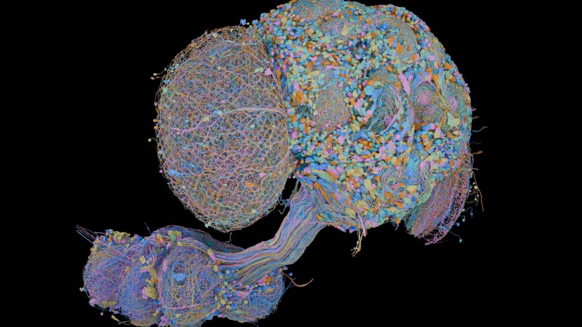
Researchers have completed a second complete fruit fly brain map, this time for an adult male’s central nervous system. The resource matters because it pairs detailed male wiring data with earlier female maps, giving scientists a way to examine where neural circuits differ between the sexes.
The project, announced September 3 by the Howard Hughes Medical Institute’s Janelia Research Campus and Google Research, was published in Cell. It maps the male fly’s brain and ventral nerve cord, rather than the brain alone, and includes more than 166,000 neurons and 125 million synaptic connections, according to Google Research.
What does the new fruit fly map cover?
A connectome is a neuron-by-neuron map of a nervous system that records synapses, the junctions through which neurons communicate. It is a wiring diagram, not a recording of brain activity or a complete account of how an animal behaves.
The new dataset covers the brain and ventral nerve cord, a structure comparable in role to a spinal cord. Including the nerve cord helps connect sensory inputs to motor outputs and gives researchers a fuller view of neural pathways involved in actions such as movement, according to Google Research and HHMI.
The term “second” needs some context. A FlyWire collaboration published a complete adult female brain connectome in 2024, covering nearly 140,000 neurons and more than 50 million synapses, according to the National Institutes of Health. Google Research also says a complete female brain-and-nerve-cord map was released earlier in 2026, so the new work should not be read as an unqualified second complete central-nervous-system map.
How did researchers build the connectome?
The team prepared the tissue in serial sections, captured electron-microscope images, and used computational methods to reconstruct its three-dimensional structure. Google Research said its systems used AI to turn the images into reconstructions of cells and connections, while human experts at Janelia annotated and proofread the finished map.
HHMI identified collaborators at the MRC Laboratory of Molecular Biology, the University of Cambridge and Google Research. Ars Technica reported that work from an intact brain to a complete connectome took about four years, with human feedback used to refine the algorithms that detected and classified synapses.
Why compare male and female fruit fly connectomes?
Google Research said the two sets of maps can be compared to find sex-linked differences in neural wiring, including circuits relevant to courtship and aggression. In areas that appear similar between the two flies, researchers can also begin measuring variation from one individual to another.
The map is intended as a research resource for testing how particular circuits may support behavior. HHMI says Janelia is applying lessons from the fly effort to connectome work in larval zebrafish and adult Danionella; the work may help make mapping larger nervous systems more practical, but it does not make a human brain map imminent.
This story draws on original reporting from Ars Technica.