
3D-printed spine models may aid planning for complex operations
A Semmelweis University survey found surgeons saw value in life-size spine models for difficult cases and patient explanations.
By Priya Raghavan · Science Reporter
3 min read
Life-size 3D-printed spine models could help surgeons prepare for some of the hardest spinal operations, according to a Semmelweis University study published in World Neurosurgery. The research also found that physical models may make it easier for doctors to explain complex spinal conditions and planned procedures to patients and families.
The study surveyed 41 specialists from three Hungarian spine surgery centers that Semmelweis University described as leading institutions. Researchers used questionnaires to compare surgeons’ views of 3D-printed anatomical models with conventional imaging tools, including X-rays, CT scans and MRI scans.
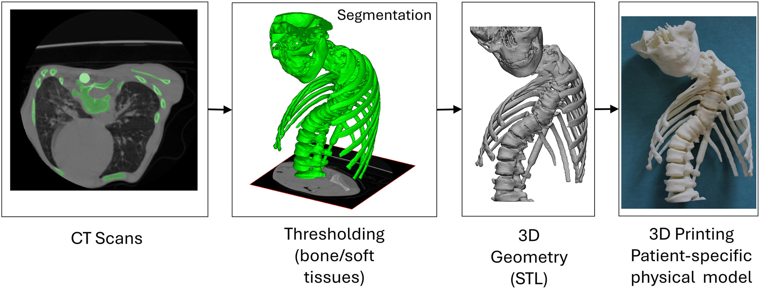
The models were made from medical imaging data and printed at life size, according to the university. The study examined cases involving congenital spinal deformities, rare developmental abnormalities, tumors and patients who had already undergone several spinal surgeries.
Where surgeons saw the most value
The researchers reported that surgeons rated the models most useful for operative planning and for communication with patients. Those advantages were strongest in cases involving unusual spinal anatomy or prior operations, including situations where metal implants made structures harder to assess on standard images.
According to Semmelweis University, the main benefit is that surgeons can study a patient-specific replica in their hands rather than relying only on images on a screen. The university said that lets clinicians view the spine from multiple angles and better assess unusual anatomy before entering the operating room.
Dr. Péter Éltes, senior author of the study, supervisor at the School of Ph.D. Studies at Semmelweis University and a spine surgeon at the National Center for Spinal Disorders, said complex anatomy can force surgeons to learn the details of each case individually. He said the models may also be used to rehearse parts of an operation, including drilling into the printed structure.
The study did not report that 3D-printed models are ready for routine clinical use. Semmelweis University said hospitals would have to meet quality assurance and regulatory requirements before such models could become a regular part of surgical practice.
Potential role in patient communication
For now, the university said the models are produced mainly for research and educational purposes. Researchers said they may later have a larger role in helping patients and relatives understand a diagnosis and a planned intervention.
Benjámin Hajnal, the study’s first author and a Ph.D. student at Semmelweis University, said a patient or parent could hold a replica of the affected spine while a physician explains the operation. He said that could be clearer than reviewing CT or MRI slices, which can be difficult for people without medical training to interpret.
The survey also found that views of the models did not vary much by surgeons’ experience level or specialty, according to the researchers. Semmelweis University said surgeons with decades of practice considered the models useful, as did younger colleagues.
The researchers reported that 80% of participating specialists had not used a 3D-printed anatomical model in clinical practice before the study. They said that finding suggests the technology may have wider potential in preparation for complex spine operations, even among surgeons who are new to the tool.
This story draws on original reporting from Medical Xpress.